

Summary: A synthetic, non-toxic form of scorpion venom helps illuminate brain tumors when stimulated by a near-infrared laser. A clinical trial of the agent showed it to be safe for use in brain cancer patients.
Source: Cedars-Sinai Medical Center
A novel imaging technique that uses a synthesized form of scorpion venom to light up brain tumors has shown promise in a clinical trial. The imaging system enables neurosurgeons to better see malignant growths that often are difficult to fully eliminate.
Results from the multi-institutional clinical trial, led by investigators from Cedars-Sinai and sponsored by Blaze Bioscience, Inc., appear in the journal Neurosurgery.
The new imaging technique that was studied uses a special high-sensitivity near-infrared camera developed at Cedars-Sinai, along with the imaging agent tozuleristide, or BLZ-100, developed by Blaze. The agent contains a synthetic version of an amino acid compound found in scorpion venom.
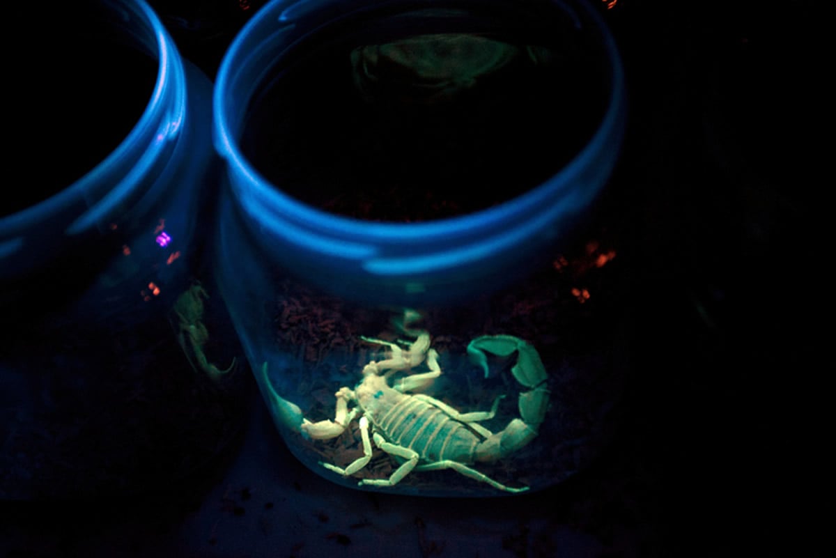
Like the natural form of the compound, the synthetic version is not toxic and binds to tumor cells. It is attached to a fluorescent dye that glows when stimulated by a near-infrared laser. Viewed through the camera, the imaging agent might allow neurosurgeons to detect the boundaries between tumors and healthy brain tissue during surgery, improving the opportunity for surgeons to remove tumor cells while sparing normal brain tissue.
“With this fluorescence, you see the tumor so much clearer because it lights up like a Christmas tree,” said Adam Mamelak, MD, senior author and investigator in the trial.
That is important because of the sprawling nature of gliomas, the type of brain tumors imaged during the trial. Gliomas are highly lethal and comprise about 33% of all brain tumors. They can infiltrate brain tissue with tentacle-like structures, making them difficult to distinguish from normal brain tissue. They typically do not respond to traditional therapies such as chemotherapy and radiation. The key to extending patient survival depends on a surgeon’s ability to detect and remove all parts of the tumor.
In the clinical trial, 17 adult patients with brain tumors were given varying doses of BLZ-100 before surgery. Despite the varying amounts of the drug given, the majority of tumors fluoresced, including both high- and low-grade gliomas. After surgery, patients were monitored for 30 days. Investigators found that none of the patients had any serious adverse responses to the drug, and that the imaging system was safe and could be useful for imaging the brain tumors during surgery.
More clinical trials are needed to further evaluate the safety of the imaging system and demonstrate the system’s effectiveness before BLZ-100 can gain approval from the Food and Drug Administration, and the camera used in the trial must be refined before it can be used seamlessly in an operating room. But Mamelak said the clinical trial results were promising.
“For a surgeon, this seamless integration of fluorescence imaging into the surgical microscope is very appealing,” Mamelak said.
Unlike other experimental systems that are bulkier or rely on multiple cameras, the new imaging system uses a single camera that takes both near-infrared and white-light images by alternating between a laser and normal white lights at very high speeds. This technology enables surgeons to easily switch back and forth between “normal” vision using a surgical microscope and fluorescent “super-vision” on a nearby monitor, in real time.
The next phase of this research, already underway, is a clinical trial involving pediatric brain tumors, taking place at up to 14 sites nationwide. This trial will serve as a data set for potential FDA approval. A similar adult clinical trial is also being planned. Although Mamelak is not directly involved in performing research during this phase, he and others are eager to see if the imaging approach has applications beyond neurosurgery.
“The technique in this study holds great promise not only for brain tumors but for many other cancer types in which we need to identify the margins of cancers,” said Keith L. Black, MD, chair of the Department of Neurosurgery at Cedars-Sinai. “The ultimate goal is to bring greater precision to the surgical care we provide to our patients.”
Discolsure Pramod Butte, MBBS, PhD and Adam Mamelak, MD are consultants for Blaze Bioscience, Inc. Pramod Butte, MBBS, PhD; Keith Black, MD and Adam Mamelak, MD are shareholders of Blaze Bioscience, Inc.
Source:
Cedars-Sinai Medical Center
Media Contacts:
Sarah R. Lichtman – Cedars-Sinai Medical Center
Image Source:
The image is adapted from the Cedars-Sinai news release.
Original Research: Closed access
“Phase 1 Safety, Pharmacokinetics, and Fluorescence Imaging Study of Tozuleristide (BLZ-100) in Adults With Newly Diagnosed or Recurrent Gliomas”. Adam Mamelak et al.
Neurosurgery. doi:10.1093/neuros/nyz125
Abstract
Phase 1 Safety, Pharmacokinetics, and Fluorescence Imaging Study of Tozuleristide (BLZ-100) in Adults With Newly Diagnosed or Recurrent Gliomas
BACKGROUND
Fluorescence-guided surgery (FGS) can improve extent of resection in gliomas. Tozuleristide (BLZ-100), a near-infrared imaging agent composed of the peptide chlorotoxin and a near-infrared fluorophore indocyanine green, is a candidate molecule for FGS of glioma and other tumor types.
OBJECTIVE
To perform a phase 1 dose-escalation study to characterize the safety, pharmacokinetics, and fluorescence imaging of tozuleristide in adults with suspected glioma.
METHODS
Patients received a single intravenous dose of tozuleristide 3 to 29 h before surgery. Fluorescence images of tumor and cavity in Situ before and after resection and of excised tissue ex Vivo were acquired, along with safety and pharmacokinetic measures.
RESULTS
A total of 17 subjects received doses between 3 and 30 mg. No dose-limiting toxicity was observed, and no reported adverse events were considered related to tozuleristide. At doses of 9 mg and above, the terminal serum half-life for tozuleristide was approximately 30 min. Fluorescence signal was detected in both high- and low-grade glial tumors, with high-grade tumors generally showing greater fluorescence intensity compared to lower grade tumors. In high-grade tumors, signal intensity increased with increased dose levels of tozuleristide, regardless of the time of dosing relative to surgery.
CONCLUSION
These results support the safety of tozuleristide at doses up to 30 mg and suggest that tozuleristide imaging may be useful for FGS of gliomas.


