Summary: A new study has upended the long-held neurological assumption that sodium concentrations are uniform across the brain’s star-shaped glial cells, or astrocytes. Researchers developed a novel imaging technique to visualize sodium content in real-time within astrocytes and their ultra-fine microscopic processes for the first time.
The data reveals that instead of a static baseline, highly specialized sodium micro-domains fluctuate dynamically across individual cells and sub-domains to match the local excitability needs of neighboring neural networks.
Key Facts
- The Glial Framework: Glial cells, including star-shaped astrocytes, comprise roughly half of the human brain. They dictate brain development, mediate communication between neurons, and regulate the excitability and functionality of neural networks.
- The Electrolyte Balance: Positively charged sodium ions (Na+), primarily sourced from dietary table salt, are the body’s most critical electrolytes. In astrocytes, maintaining a low internal sodium concentration is mandatory for regulating neurotransmitters at synaptic junctions and balancing secondary electrolytes.
- Dismantling the Uniformity Baseline: Neurobiologists long assumed that sodium concentrations remained at an identical, uniformly low baseline across all astrocytes and their cellular sub-units to ensure reliable housekeeping. The team’s new direct-tissue imaging method explicitly disproved this, capturing stark baseline variations between individual cells and within their sub-structures.
- Membrane Transport Architectures: Collaborating with researchers at Friedrich-Alexander-Universität Erlangen-Nuremberg, the team proved that these localized sodium differences are driven by specific transport molecules, which present in shifting numbers and structural configurations across various astrocyte membranes.
- Multi-Scale Validation Framework: The experimental findings gathered from isolated brain tissue at HHU were integrated into biophysical computer simulations by the University of South Florida and fully validated in living animal models by the University of Bonn and University Hospital Bonn.
- Clinical Disruptions: Lead investigators note that these newly mapped sodium sub-domains react dynamically to neighboring synaptic needs. Consequently, failures in these localized electrolyte balances provide essential research targets for neurological disorders where ion regulation collapses, such as epilepsy or acute stroke.
Source: HHU
The brain does not solely comprise nerve cells (neurons); roughly half of the organ is made up of so-called glial cells, which play an important role in brain development and are crucial for communication between neurons and the function of neural networks. Glial cells also include so-called star cells or “astrocytes”.
The element sodium, or rather positively charged sodium ions, are the most important electrolytes in the human body. These ions are crucial for many bodily functions. The main source thereof is table salt (NaCl), which is obtained from food.
Sodium ions are also involved in many processes in the brain, meaning that their concentration must be strictly regulated. In astrocytes, a low intracellular sodium concentration is important among other things for the regulation of neurotransmitters at the synapses – the junctions between nerve cells. It is also important for regulating the levels of other electrolytes. This enables astrocytes to ensure the functionality of nerve cells and regulate their excitability.
At the Institute of Neurobiology at HHU, the team led by Professor Dr Christine Rose has now developed a new technique as part of a study (the SynGluCross project) funded by the Federal Ministry of Education and Research (Bundesministerium für Bildung und Forschung – BMBF), which can make the sodium content in the astrocytes and their fine processes directly visible in brain tissue for the first time.
Together with researchers from Friedrich-Alexander-Universität Erlangen-Nuremberg, the University of Bonn, the University Hospital Bonn, and the University of South Florida in Tampa (USA), the neurobiologists in Düsseldorf set out to test the existing assumption that there is a similarly low concentration of sodium in all astrocytes and in all their sub-units to enable the astrocytes to perform their vital tasks reliably.
They actually established that this is not the case. Rather, they discovered differences – both between individual astrocytes and within various sub-units of these cells. Together with their colleagues from Erlangen-Nuremberg, they also demonstrated that certain transport molecules, which can be found in the cell membrane of various astrocytes in differing numbers and configurations, are responsible for these differences.
The cooperation partners from the USA implemented these findings in biophysical computer models and were able to replicate the experimental results in simulations. The findings obtained in isolated brain tissue in Düsseldorf were validated in animal models by the colleagues in Bonn.
Dr Jan Meyer, lead author of the study: “We were also able to show that specialised functional sub-domains exist in astrocytes due to the different sodium concentrations. In each case, they react to the local needs of their neighbouring neural network.”
The head of the study, Professor Christine Rose, highlights further aspects: “These newly discovered properties of astrocytes may also play a role in various brain disorders where ion levels and neurotransmitter regulation are disrupted, such as epilepsy, or after a stroke. Our findings thus offer starting points for further research.”
Key Questions Answered:
A: To serve as a responsive neighbor. Astrocytes are responsible for managing neurotransmitters and keeping nerve cell signals under control. Because different synapses in a neural network are firing at different rates, the astrocyte creates specialized, isolated sodium sub-domains within its fine branches to instantly match the custom, hyper-local needs of nearby neurons.
A: It was a complete multi-scale validation. The primary discovery was made using a new imaging technique on isolated brain tissue in Düsseldorf. To confirm it wasn’t a laboratory anomaly, biophysicists in South Florida built computer models that perfectly mirrored the patterns in simulations, while neurobiologists in Bonn verified the exact same localized variations inside living animal models.
A: It offers a completely fresh direction for targeted drug research. Conditions like epilepsy and stroke are fundamentally driven by massive, toxic disruptions in brain ion levels and neurotransmitter regulation. Knowing that astrocytes rely on specific transport molecules to manage these hyper-local sodium sub-domains allows scientists to develop medications that protect these cellular pumps from collapsing during an emergency.
Editorial Notes:
- This article was edited by a Neuroscience News editor.
- Journal paper reviewed in full.
- Additional context added by our staff.
About this neuroscience research news
Author: Arne Claussen
Source: HHU
Contact: Arne Claussen – HHU
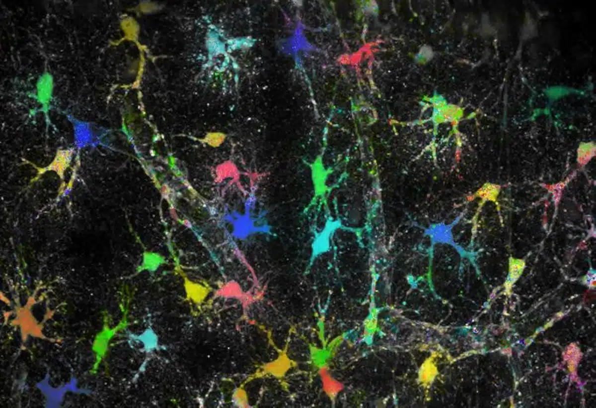
Image: The image is credited to HHU/Institute of Neurobiology – Jan Meyer
Original Research: Open access.
“Cellular and subcellular heterogeneity of astrocytic Na⁺ homeostasis tuning astrocytes into functionally distinct subgroups in the mouse brain” by Jan Meyer, Viola Bornemann, Alok Bhattarai, Sara Eitelmann, Petr Unichenko, Simone Durry, Karl W. Kafitz, Nicholas Chalmers, Jianfeng Fan, Ruth Beckervordersandforth, Christian Henneberger, Ghanim Ullah & Christine R. Rose. Nature Communications
DOI:10.1038/s41467-026-73435-z
Abstract
Cellular and subcellular heterogeneity of astrocytic Na⁺ homeostasis tuning astrocytes into functionally distinct subgroups in the mouse brain
Astrocytes maintain extracellular ion and transmitter homeostasis, with the Na⁺ inward gradient playing a crucial role. Earlier studies suggested a rather low, uniform Na⁺ distribution in astrocytes, consistent with the view that these basic homeostatic properties are well-protected.
Here, we employed multi-photon fluorescence lifetime imaging to quantitatively determine astrocytic [Na+] in mouse brain tissue slices and in vivo.
Our data reveals a significant subcellular and cellular heterogeneity in astrocytic [Na+], accompanied by differences in the capacity for Na+/K+-ATPase (NKA)-mediated uptake of extracellular K+.
RNAscope and immunohistochemistry indicate differential spatial expression patterns of NKA ß1 and ß2 subunits in astrocytes. Biophysical modeling of differential NKA expression together with varying strength of Na+ influx replicate the experimentally observed heterogeneity in astrocytic [Na+].
Altogether, our results suggest the existence of functionally distinct astrocytes and astrocyte subdomains in which Na+ homeostasis is locally adapted to the specific requirements of surrounding neural networks.