New Insights Into Activity Patterns Inside the Anesthetized Brain

Summary: Under anesthesia, the nerve cells change their mode of operation by firing more synchronously, and by becoming surprisingly reactive to environmental stimuli, researchers report.

Source: Charite.

Even under deep anesthesia, nerve cells remain highly active. A study conducted by researchers from Charité – Universitätsmedizin Berlin has shown by high-resolution cellular imaging that local neuronal networks remain active even when the brain is unconscious. Under anesthesia, the nerve cells change their mode of operation by firing more synchronously, and by becoming surprisingly reactive to environmental stimuli. Results from this research have been published in the journal Frontiers in Cellular Neuroscience.

Establishing how the brain produces consciousness is one of the most challenging research questions in the field of neuroscience. In an effort to get closer to an answer, a team led by Dr. Mazahir T. Hasan, a researcher with Charité’s NeuroCure Cluster of Excellence, joined forces with colleagues from the Max Planck Institute for Medical Research in Heidelberg. By visualizing neuronal activity in the brains of mice, they were able to compare how brain activity differs in conscious and anesthetized mice. NeuroCure’s Dr. Hasan explains: “We used a fluorescent protein that converts electrical signals into light signals. This enabled us to visualize the frequency and average amplitude of neuronal responses, and allowed us to reveal the existence of neuronal synchrony.” Results from this research would suggest that consciousness is not simply dependent on the number of active neurons inside the cortex; instead, it seems to be dependent upon the way these nerve cells communicate and on the degree to which they manage to differ in terms of their activity patterns.

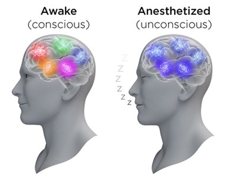
The awake cortex showed complex activity patterns, with individual cells firing at different times. Under anesthesia, all neurons displayed identical activity patterns and fired at the same time. “While one might expect the brain to cease its activity under anesthesia, in reality, the situation is quite different. Neurons remain highly active but change their communication mode. During unconsciousness they become highly synchonized – in simple terms all neurons start doing the same thing. ” explains Mr. Thomas Lissek, a neurobiologist from Heidelberg and the study’s first author. Another surprising finding was that neurons were more sensitive to environmental stimuli under anesthesia than when the brain was awake. “This is especially surprising, as anesthesia is used to block both pain and environmental stimuli during surgery,” says Mr. Lissek. Some of the brain regions that are normally dedicated to tactile perception even responded to sound information.

Image shows brain activity.
Highly active and synchronized: nerve cells under anesthesia. Neurosciencenews image is credited to Thomas Splettstoesser.

These new insights into neuronal activity patterns provide information regarding the identity of the cellular parameters involved in producing consciousness and the loss of consciousness. Once combined with further advances that would allow us to measure neuronal activity inside the human brain, these findings could contribute to improving our diagnostic capabilities in conditions such as coma and locked-in syndrome. For the first time, this study succeeded in showing that it is possible to observe visually identifiable neuronal networks over a period of several weeks in order to study the after-effects of anesthesia. “It is clear that investigation of anesthesia will produce deep insights into the mechanism of consciousness”, emphasizes Hasan.

About this neuroscience research article

Source: Charite
Image Source: This NeuroscienceNews.com image is credited to Thomas Splettstoesser.
Original Research: Full open access research for “General Anesthetic Conditions Induce Network Synchrony and Disrupt Sensory Processing in the Cortex” by Thomas Lissek, Horst A. Obenhaus, Désirée A. W. Ditzel, Takeharu Nagai, Atsushi Miyawaki, Rolf Sprengel and Mazahir T. Hasan in Frontiers in Cellular Neuroscience. Published online April 14 2016 doi:10.3389/fncel.2016.00064

Cite This NeuroscienceNews.com Article

[cbtabs][cbtab title=”MLA”]Charite. “New Insights Into Activity Patterns Inside the Anesthetized Brain.” NeuroscienceNews. NeuroscienceNews, 11 November 2016.
<https://neurosciencenews.com/anesthesia-neural-activity-5494/>.[/cbtab][cbtab title=”APA”]Charite. (2016, November 11). New Insights Into Activity Patterns Inside the Anesthetized Brain. NeuroscienceNews. Retrieved November 11, 2016 from https://neurosciencenews.com/anesthesia-neural-activity-5494/[/cbtab][cbtab title=”Chicago”]Charite. “New Insights Into Activity Patterns Inside the Anesthetized Brain.” https://neurosciencenews.com/anesthesia-neural-activity-5494/ (accessed November 11, 2016).[/cbtab][/cbtabs]


Abstract

General Anesthetic Conditions Induce Network Synchrony and Disrupt Sensory Processing in the Cortex

General anesthetics are commonly used in animal models to study how sensory signals are represented in the brain. Here, we used two-photon (2P) calcium activity imaging with cellular resolution to investigate how neuronal activity in layer 2/3 of the mouse barrel cortex is modified under the influence of different concentrations of chemically distinct general anesthetics. Our results show that a high isoflurane dose induces synchrony in local neuronal networks and these cortical activity patterns closely resemble those observed in EEG recordings under deep anesthesia. Moreover, ketamine and urethane also induced similar activity patterns. While investigating the effects of deep isoflurane anesthesia on whisker and auditory evoked responses in the barrel cortex, we found that dedicated spatial regions for sensory signal processing become disrupted. We propose that our isoflurane-2P imaging paradigm can serve as an attractive model system to dissect cellular and molecular mechanisms that induce the anesthetic state, and it might also provide important insight into sleep-like brain states and consciousness.

“General Anesthetic Conditions Induce Network Synchrony and Disrupt Sensory Processing in the Cortex” by Thomas Lissek, Horst A. Obenhaus, Désirée A. W. Ditzel, Takeharu Nagai, Atsushi Miyawaki, Rolf Sprengel and Mazahir T. Hasan in Frontiers in Cellular Neuroscience. Published online April 14 2016 doi:10.3389/fncel.2016.00064

Feel free to share this Neuroscience News.
Join our Newsletter
I agree to have my personal information transferred to AWeber for Neuroscience Newsletter ( more information )
Sign up to receive our recent neuroscience headlines and summaries sent to your email once a day, totally free.
We hate spam and only use your email to contact you about newsletters. You can cancel your subscription any time.
  1. Anaesthesia is not the result of simply turning neural circuits off as was originally believed. The process of becoming anaesthetised with some agents may, paradoxically, show increased brain activity in some brain areas as is discussed in the discussion section of this excellent paper. The paradoxical finding of increased sensory activity under anaesthesia may occur in a similar manner to increased neurone activity seen in hyperacusis during induction of anaesthesia (and with the development of tinnitus). This really interesting research may also offer an explanation for the development of emergence of postoperative delirium (POD). The normal vigilance circuits of the amygdala are no longer under the ameliorating influence of the neocortex as the latter is more inhibited by anaesthetics than the former. Any associative learning (memory) between normal non noxious stimuli during anaesthesia and noxious stimuli (surgery) may leave a legacy that emerges in the postoperative period as patients emerge into a similar ambience. Essentially similar to “amygdala hijack”. What do you think?

  2. This finding suggests (to me) that when anesthetized (or a sleep) a neural Actention Selection Serving System is largely taking a break.

    An individual ASSS selects what to respond to among all internal and external sources of sensory-motivational messages.

    [More exhaustively put: an ASSS does so according to past and current influences environmentally presented to it – and also according to epigenetically and phylogenetically significant naturally selective factors operating on top of by quantum reality ‘preordained’ ;> (hence most intrinsic possible) evolutionary patterning trend effecting probabilistic type potentials.]

    How it does so fairly obviously involves a very intensive and of course multi-threaded/faceted process — one involving mostly subliminally active, adaptively/functionally incompatible and hence mutually but seldom equally competing, actention modules. That is, such actention modules as if compete with each other by exerting direct inhibitory, or via interneurons inhibitory, influences on each other.

    This way, unless the nocturnal sleep or seasonally active aestivation module or hiberantion module or another to adaptive seasonal dormancy dedicated module is dominant within the ASSS, it is possible for one or the other of our mutually incompatible actention modules to for some limited period of time become a “focused” and “paid” (primarily in the currency of neurometabolic resources) actention module.

    The most central arena for the competition between adaptively/functionally incompatible actention modules (in our brains) might be the basal ganglia.

    P.S.
    “AS” the shortest accEPTable alternative to writing “ASSS”. Neither spelling version is the most nor the least SEPTIC humored concEPT expressible as a fun and boredom/botheredness-relieving acronym; And even if the intermediate-length spelling is used it would be neither the most nor the least SEPTIC humored of the only 3 such concEPT. I have placed the AS-version right in the middle of the most integrative of all evolutionary psychology type concEPTs (i.e. concepts at least superficially unique to my MAD-inspired “ÆPT” worldview and only fairly (e.g. not financially) philanthropically oriented outlook on/understanding of ourselves.

    I was referring to “EAVASIVE”. EAVASIVE is hardly at all a humorous concEPT.

    The least SEPTIC humored of the 3 thusly jocular concepts is “RAT (reticular activating type) neurons”.

    The firing of RAT neurons is the only firmly known ‘functural’ factor found to be an essential for any level (mode/mood/intensity/content) of consciousness – or of ‘paying actention’.

    Our actention modules are competing in face of all significant parallel situational challenges.

    It is of central ÆPT concern that, very often, challenges of opportunity type parallel or overlap with current environmentally presented threats of “Specific/synaptic Hibernation {SH, for short, refers to a highly specific post-synaptically metabolism-muting function} Imploring Type”;

    And, that, even more often, environmentally present opportunities overlap with through “operant conditioning” left behind (dynamic imprints of) past “SHI type” threats;

    And, that current SHI type challenges combine with previously accumulated CURSES and also with opportunity type challenges.

    EAVASIVE encapsulates precisely that (and it implies some Darwinian evolutionary pressure type specifics — included in ÆPT — of how) some particular occasions of momentous mutations improved our common ancestral individuals’ capacity to cope with such threats while also improving the capability of exploiting concurrent opportunity type challenges (i.e. more or less directly procreation promoting – if exploited or taken – environmentally present opportunities).

    These (by SHI type threats) imprinted states (neural imprints of a primarily insidious character) are kept more or less adaptively sequestered by by synapse blocking functions but also to a uniquely human extent also as if recycled (advantageously rerouted) by “synapse sprouting” functions.

    Even in most post traumatic (post-SHIT) circumstances it would be maladaptive if the automatically imprinted aftermath of such threats started to be belatedly responded to.
    However, in a sense, it is also ÆPT to describe such an imprint as an indefinitely postponed response to a SHI type threat; That is, an originally to have been maladaptive responses (actentions) that became postponed at least until an ideal enough lifetime situation may arise so that it would be possible to actively reprocess and thus adaptively respond (actend) to the as if neurally preserved original ordeal (of SHI type).

    Any single such “aftermath” (of a SHI type threat) suits being referred to as a “Conditioned-in kept Unconscious Reverberating State Effecting Symptoms” – or, by tersest possible abbreviation, a “CURSES”.

    PPS
    I hope you appreciate that I have rather successfully — partly by using a most distinKt example of SEPTIC humor — relieved myself of almost inevitably having had to use the tacky Freudian word “trauma”. ;-)

Comments are closed.