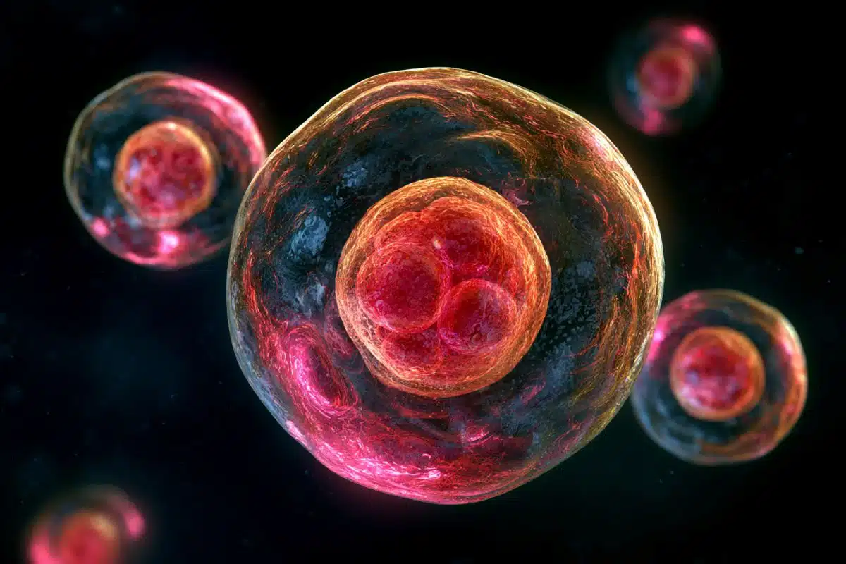
Summary: Amyloid precursor protein accumulated with an excess of presynaptic proteins, whereas post synaptic proteins were depleted.
Source: Wyss Center for Bio and Neuroengineering
New research published today in Alzheimer’s & Dementia: The Journal of the Alzheimer’s Association could explain why neurons fail to communicate effectively in people with Alzheimer’s disease (AD).
The study conducted by researchers in the laboratory of Christophe Mulle at the Interdisciplinary Institute of Neuroscience in Bordeaux1 opens a new research path to establish the molecular mechanism causing AD.
Despite intense efforts in clinical research, there is no treatment to cure or even slow down the progression of AD. Current clinical interventions are typically based on the ‘amyloid cascade’ hypothesis, postulating that neurodegeneration in AD is caused by the abnormal accumulation of amyloid plaques in the brain.
However, these interventions have failed to demonstrate clinical efficacy. Recently, the European Medicines Agency did not approve a controversial new drug for AD that targets plaques, concluding that the benefits do not outweigh the risks. The decision leaves an estimated 8 million people living with dementia in the EU with no treatment options, highlighting the urgent need for an alternative target for AD research.
The new study reveals that the amyloid peptide composing the plaques is not the only offender accumulating in the human AD brain. Its precursor, the amyloid precursor protein (APP), was found to surround amyloid plaques in intense microscopic staining.
“Remarkably, our research revealed that the areas of the brain where APP accumulates, contain abnormal amounts of proteins essential for the communication between neurons,” said Dr Gael Barthet senior author on the paper, now Director of Neuroscience at the Wyss Center.
The communication between neurons occurs at junctions called synapses where neurotransmitters released by a pre-synaptic neuron cross the junction to activate a post-synaptic neuron.

“What we found fascinating was that APP accumulated with excess of presynaptic proteins whereas post-synaptic proteins where depleted, indicating severe impairment of neuronal communication at these sites,” said Dr Tomas Jorda, lead author of the paper and Post-Doctoral Research Associate in neurobiology at the University of Geneva.
“The new findings introduce a new direction for us to explore using our innovative, multi-scale, multi-modal approach at the Center,” said Dr Richie Kohman, Chief Scientific Officer at the Wyss Center.
The Wyss Center’s translational research on neurodegenerative diseases uses novel molecular labelling techniques coupled with advanced microscopy and data analysis tools to examine specific neuronal populations, synaptic markers, and single-cell transcriptomics in the human brain. These innovative imaging approaches enable the study of the human brain in large sections or even in entire 3D samples. The Wyss Center is using this pipeline to investigate the molecular mechanisms at play in neurological and psychiatric disorders, to establish early biomarkers and identify new therapeutic targets.
About this Alzheimer’s disease research news
Author: Jo Bowler
Source: Wyss Center for Bio and Neuroengineering
Contact: Jo Bowler – Wyss Center for Bio and Neuroengineering
Image: The image is credited to Gael Barthet
Original Research: Open access.
“APP accumulates with presynaptic proteins around amyloid plaques: A role for presynaptic mechanisms in Alzheimer’s disease?” by Gael Barthet et al. Alzheimer’s and Dementia
Abstract
APP accumulates with presynaptic proteins around amyloid plaques: A role for presynaptic mechanisms in Alzheimer’s disease?
In Alzheimer’s disease (AD), the distribution of the amyloid precursor protein (APP) and its fragments other than amyloid beta, has not been fully characterized.
Here, we investigate the distribution of APP and its fragments in human AD brain samples and in mouse models of AD in reference to its proteases, synaptic proteins, and histopathological features characteristic of the AD brain, by combining an extensive set of histological and analytical tools.
We report that the prominent somatic distribution of APP observed in control patients remarkably vanishes in human AD patients to the benefit of dense accumulations of extra-somatic APP, which surround dense-core amyloid plaques enriched in APP-Nter. These features are accentuated in patients with familial forms of the disease.
Importantly, APP accumulations are enriched in phosphorylated tau and presynaptic proteins whereas they are depleted of post-synaptic proteins suggesting that the extra-somatic accumulations of APP are of presynaptic origin. Ultrastructural analyses unveil that APP concentrates in autophagosomes and in multivesicular bodies together with presynaptic vesicle proteins.
Altogether, alteration of APP distribution and its accumulation together with presynaptic proteins around dense-core amyloid plaques is a key histopathological feature in AD, lending support to the notion that presynaptic failure is a strong physiopathological component of AD.