Summary: Researchers apply deep learning algorithms to help measure physiological changes linked to heart disease.
Source: USC.
Heart disease is the leading cause of death for both men and women, according to the Centers for Disease Control and Prevention (CDC). In the U.S., one in every four deaths is a result of heart disease, which includes a range of conditions from arrhythmias, or abnormal heart rhythms, to defects, as well as blood vessel diseases, more commonly known as cardiovascular diseases.
Predicting and monitoring cardiovascular disease is often expensive and tenuous, involving high-tech equipment and intrusive procedures. However, a new method developed by researchers at USC Viterbi School of Engineering offers a better way. By coupling a machine learning model with a patient’s pulse data, they are able to measure a key risk factor for cardiovascular diseases and arterial stiffness, using just a smart phone.
Arterial stiffening, in which arteries become less elastic and more rigid, can result in increased blood and pulse pressure. In addition to being a known risk factor for cardiovascular diseases, it is also associated with diseases like diabetes and renal failure.
“If the aorta is stiff, then when it transfers the pulse energy all the way to the peripheral vasculature – to small vessels – it can cause end organ damage. So, if the kidneys are sitting at the end, the kidneys get hurt; if the brain is sitting at the end, the brain gets hurt,” said Niema Pahlevan, assistant professor of aerospace and mechanical engineering and medicine.
Checking for a pulse
By measuring pulse wave velocity, which is the speed that the arterial pulse propagates through the circulatory system, clinicians are able to determine arterial stiffness. Current measurement methods include MRI, which is expensive and often not feasible, or tonometry, which requires two pressure measurements and an electrocardiogram to match the phases of the two pressure waves.
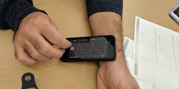
The novel method developed by Pahlevan, Marianne Razavi and Peyman Tavallali uses a single, uncalibrated carotid pressure wave that can be captured with a smart phone’s camera. In a previous study, the team used the same technology to develop an iPhone app that can detect heart failure using the slight perturbations of your pulse beneath your skin to record a pulse wave. In the same fashion, they are able to determine arterial stiffness.
“An uncalibrated, single waveform – that means that you eliminated two steps. That’s how you go from an $18,000 tonometry device and intrusive procedure to an iPhone app,” Pahlevan said.
“It’s very easy to operate,” added Razavi, who is the director of biostatistics for Avicena LLC, the startup company developing the app. “I actually taught my kid to do it.”
Instead of a detailed waveform required with tonometry, their method needs just the shape of a patient’s pulse wave for the mathematical model, called intrinsic frequency, to calculate key variables related to the phases of the patient’s heartbeat. These variables are then used in a machine learning model that determines pulse wave velocity (PWV) and, therefore, arterial stiffness.
Validation
To validate their method, they used existing tonometry data collected from the Framingham Heart Study, a long-term epidemiological cohort analysis. Using 5,012 patients, they calculated their own PWV measurements and compared them with the tonometry measurements from the study, finding an 85 percent correlation between the two.
But more importantly, they needed to determine whether their method could be used to predict cardiovascular disease.
“What the clinician wants to know is whether or not you’re helping them to improve outcome,” Pahlevan said. “And we showed that it is as predictive as the actual tonometry.”
Through a prospective study using 4,798 patients, they showed that their PWV measurement was significantly associated with the onset of cardiovascular diseases over a ten-year follow up period. Their study was published in Nature Scientific Reports in January.
Bringing AI to medicine
“A lot of people have tried to bring machine learning to medical devices, but pure AI by itself doesn’t work,” Pahlevan said. “When you get a high correlation, you can be missing all of the diseased patients because, in medicine, the outliers are the cases you want to capture – they’re the important ones.”
The reason their machine learning method is able to capture clinically significant outcomes is due to their intrinsic frequency algorithm, which is the mathematical analysis used to calculate physically relevant variables relating to the patient’s heart and vascular function. The main variables represent the heart’s performance during the contraction phase (systole) and the vasculature’s performance during the relaxed phase (diastole).
Developed just three years ago during Pahlevan’s postdoctoral work, the team plans on expanding on the intrinsic frequency algorithm so that it can be applied to a number of other applications, such as detecting silent heart attacks.
Source: USC
Publisher: Organized by NeuroscienceNews.com.
Image Source: NeuroscienceNews.com image is credited to Ashleen Knutsen.
Original Research: Open access research for “Artificial Intelligence Estimation of Carotid-Femoral Pulse Wave Velocity using Carotid Waveform” by Peyman Tavallali, Marianne Razavi & Niema M. Pahlevan in Scientific Reports. Published January 27 2018.
doi:10.1038/s41598-018-19457-0
[cbtabs][cbtab title=”MLA”]USC “Using AI to Detect Heart Disease.” NeuroscienceNews. NeuroscienceNews, 17 April 2018.
<https://neurosciencenews.com/ai-heart-disease-8825/>.[/cbtab][cbtab title=”APA”]USC (2018, April 17). Using AI to Detect Heart Disease. NeuroscienceNews. Retrieved April 17, 2018 from https://neurosciencenews.com/ai-heart-disease-8825/[/cbtab][cbtab title=”Chicago”]USC “Using AI to Detect Heart Disease.” https://neurosciencenews.com/ai-heart-disease-8825/ (accessed April 17, 2018).[/cbtab][/cbtabs]
Abstract
Artificial Intelligence Estimation of Carotid-Femoral Pulse Wave Velocity using Carotid Waveform
In this article, we offer an artificial intelligence method to estimate the carotid-femoral Pulse Wave Velocity (PWV) non-invasively from one uncalibrated carotid waveform measured by tonometry and few routine clinical variables. Since the signal processing inputs to this machine learning algorithm are sensor agnostic, the presented method can accompany any medical instrument that provides a calibrated or uncalibrated carotid pressure waveform. Our results show that, for an unseen hold back test set population in the age range of 20 to 69, our model can estimate PWV with a Root-Mean-Square Error (RMSE) of 1.12 m/sec compared to the reference method. The results convey the fact that this model is a reliable surrogate of PWV. Our study also showed that estimated PWV was significantly associated with an increased risk of CVDs.