

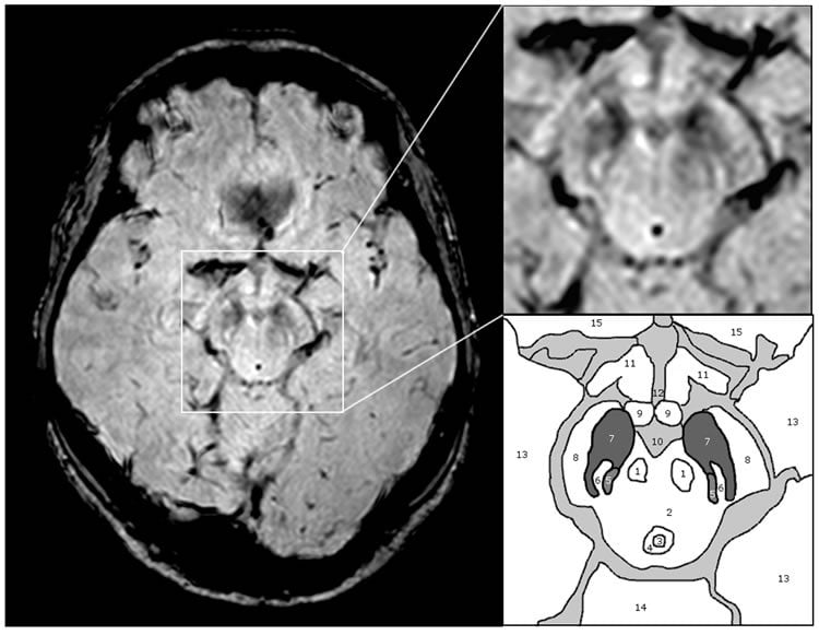
An image similar in shape to a Swallow’s tail has been identified as a new and accurate test for Parkinson’s disease. The image, which depicts the healthy state of a group of cells in the sub-region of the human brain, was singled out using 3T MRI scanning technology – standard equipment in clinical settings today.
The research was led by Dr Stefan Schwarz and Professor Dorothee Auer, experts in neuroradiology in the School of Medicine at The University of Nottingham and was carried out at the Queen’s Medical Centre in collaboration with Dr Nin Bajaj, an expert in Movement Disorder Diseases at the Nottingham University Hospitals NHS Trust.
The findings have been published in the open access academic journal PLOS ONE.
The work builds on a successful collaboration with Professor Penny Gowland at the Sir Peter Mansfield Magnetic Resonance Centre at The University of Nottingham.
‘The ‘Swallow Tail’ Appearance of the Healthy Nigrosome – A New Accurate Test of Parkinson’s Disease: A Case-Control and Retrospective Cross-Sectional MRI Study at 3T’ – describes how the absence of this imaging sign can help to diagnose Parkinson’s disease using standard clinical Magnetic Resonance Scanners.
Parkinson’s disease is a progressive neurodegenerative disorder which destroys brain cells that control movement. Around 127,000 people in the UK have the disease. Currently there is no cure but drugs and treatments can be taken to manage the symptoms.
The challenges of diagnosing Parkinson’s
Until now diagnosing Parkinson’s in clinically uncertain cases has been limited to expensive nuclear medical techniques. The diagnosis can be challenging early in the course of the condition and in tremor dominant cases. Other non-licensed diagnostic techniques offer a varying range of accuracy, repeatability and reliability but none of them have demonstrated the required accuracy and ease of use to allow translation into standard clinical practice.
Using high resolution, ultra high filed 7T magnetic resonance imaging the Nottingham research team has already pinpointed the characteristic pathology of Parkinson’s with structural change in a small area of the mid brain known as the substantia nigra. The latest study has shown that these changes can also be detected using 3T MRI technology which is accessible in hospitals across the country. They subsequently coined the phrase the ‘swallow tail appearance’ as an easy recognizable sign of the healthy appearing substantia nigra which is lost in Parkinson’s disease. A total of 114 high-resolution scans were reviewed and in 94 per cent of cases the diagnosis was accurately made using this technique.
New findings give new hope
Dr Schwarz said: “This is a breakthrough finding as currently Parkinson’s disease is mostly diagnosed by identifying symptoms like stiffness and tremor. Imaging tests to confirm the diagnosis are limited to expensive nuclear medical techniques which are not widely available and associated with potentially harmful ionizing radiation.
“Using Magnetic Resonance Imaging (no ionizing radiation involved and much cheaper than nuclear medical techniques) we identified a specific imaging feature which has great similarity to a tail of a swallow and therefore decided to call it the ‘swallow tail sign’. This sign is absent in Parkinson’s disease.”
The research was funded by The University of Nottingham, the Sarah Matheson Trust, and the Medical Research Council. Dr Schwarz’s Academic Clinical Lectureship is funded by the National Institute for Health Research UK.
The funders had no role in study design, data collection and analysis.
Contact: Lindsay Brooke – University of Nottingham
Source: University of Nottingham press release
Image Source: The image is credited to Schwarz et al./PLOS ONE and is adapted from the open access research paper
Original Research: Full open access research for “The ‘Swallow Tail’ Appearance of the Healthy Nigrosome – A New Accurate Test of Parkinson’s Disease: A Case-Control and Retrospective Cross-Sectional MRI Study at 3T” by Stefan T. Schwarz, Mohammed Afzal, Paul S. Morgan, Nin Bajaj, Penny A. Gowland, and Dorothee P. Auer in PLOS ONE. Published online April 7 2014 doi:10.1371/journal.pone.0093814
The ‘Swallow Tail’ Appearance of the Healthy Nigrosome – A New Accurate Test of Parkinson’s Disease: A Case-Control and Retrospective Cross-Sectional MRI Study at 3T
There is no well-established in vivo marker of nigral degeneration in Parkinson’s disease (PD). An ideal imaging marker would directly mirror the loss of substantia nigra dopaminergic neurones, which is most prominent in sub-regions called nigrosomes. High-resolution, iron-sensitive, magnetic resonance imaging (MRI) at 7T allows direct nigrosome-1 visualisation in healthy people but not in PD. Here, we investigated the feasibility of nigrosome-1 detection using 3T – susceptibility-weighted (SWI) MRI and the diagnostic accuracy that can be achieved for diagnosing PD in a clinical population. 114 high-resolution 3T – SWI-scans were reviewed consisting of a prospective case-control study in 19 subjects (10 PD, 9 controls) and a retrospective cross-sectional study in 95 consecutive patients undergoing routine clinical SWI-scans (>50 years, 9 PD, 81 non-PD, 5 non-diagnostic studies excluded). Two raters independently classified subjects into PD and non-PD according to absence or presence of nigrosome-1, followed by consensus reading. Diagnostic accuracy was assessed against clinical diagnosis as gold standard. Absolute inter- and intra-rater agreement was ≥94% (kappa≥0.82, p<0.001). In the prospective study 8/9 control and 8/10 PD; and in the retrospective study 77/81 non-PD and all 9 PD subjects were correctly classified. Diagnostic accuracy of the retrospective cohort was: sensitivity 100%, specificity 95%, NPV 1, PPV 0.69 and accuracy 96% which dropped to 91% when including non-diagnostic scans (‘intent to diagnose’). The healthy nigrosome-1 can be readily depicted on high-resolution 3T – SWI giving rise to a ‘swallow tail’ appearance of the dorsolateral substantia nigra, and this feature is lost in PD. Visual radiological assessment yielded a high diagnostic accuracy for PD vs. an unselected clinical control population. Assessing the substantia nigra on SWI for the typical ‘swallow tail’ appearance has potential to become a new and easy applicable 3T MRI diagnostic tool for nigral degeneration in PD.
“The ‘Swallow Tail’ Appearance of the Healthy Nigrosome – A New Accurate Test of Parkinson’s Disease: A Case-Control and Retrospective Cross-Sectional MRI Study at 3T” by Stefan T. Schwarz, Mohammed Afzal, Paul S. Morgan, Nin Bajaj, Penny A. Gowland, and Dorothee P. Auer in PLOS ONE, April 7 2014 doi:10.1371/journal.pone.0093814


