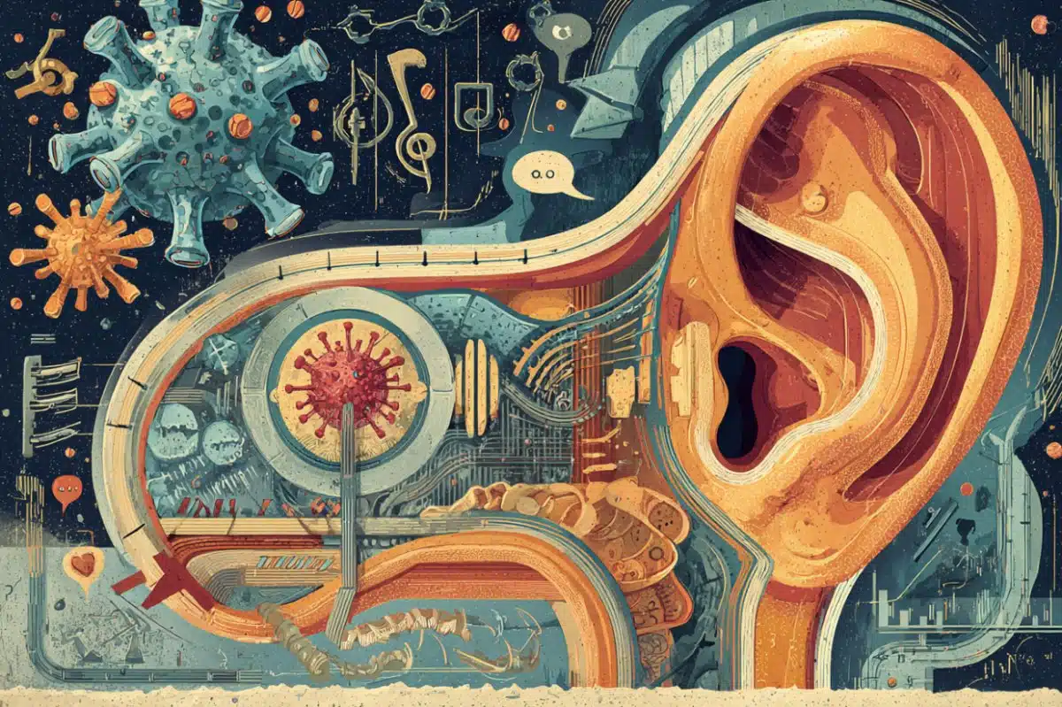
New study finds that blockages in fruit fly brains quickly form and dissolve; it could help treat Alzheimer’s and Huntington’s diseases.
Motorists in Los Angeles, San Francisco and other gridlocked cities could learn something from the fruit fly.
Scientists have found that cellular blockages, the molecular equivalent to traffic jams, in nerve cells of the insect’s brain can form and dissolve in 30 seconds or less.
The findings, presented in the journal PLOS ONE, could provide scientists much-needed clues to better identify and treat neurodegenerative diseases such as Alzheimer’s and Huntington’s.
“Our research suggests that fixed, permanent blocks may impede the transport of important cellular components and, ultimately, lead to cellular degeneration and death,” says lead researcher Shermali Gunawardena, PhD, an assistant professor of biological sciences in the University at Buffalo’s College of Arts and Sciences. “Conversely, blocks that resolve themselves may be benign.”
She continues: “This is an important distinction that could help researchers decide which kind or type of blocks to focus on when developing drugs and other forms of therapy for some of these debilitating diseases.”
Scientists have long known that many essential cellular components are transported along tracts of nerve cells called neuronal pathways, and that these movements are required for the growth, function and maintenance of neurons. Only recently, however, have they been able to understand the many proteins that help control these movements.
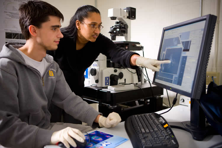
In the UB study, researchers examined isolated nerve cells from fruit fly larvae. Neuronal pathways of these larvae are similar to neuronal pathways in humans.
Traditionally, researchers have identified blockages through still images of dead larvae. These images provide a snapshot only, instead of a depiction of the behavior of the accumulated components over distinct periods of time.
UB researchers altered the approach by analyzing the neuronal pathways of living larvae. Unlike the still images, this method shows how the transport of components changes as neuronal pathways evolve over time.
The researchers found that certain blockages form and dissolve rather quickly. For example, one blockage appeared and disappeared within 29 seconds. Its relatively short life, Gunawardena said, indicates that the blockage is likely benign and not harmful to the cell.
Research by UB biologists suggests that blockages in fruit fly brains that form and dissolve quickly may be benign, a finding that could have implications for the treatment of diseases like Alzheimer’s and Huntington’s in people.
The distinction is significant, she said, because it could allow researchers to focus on permanent blockages that likely halt cellular movement and may pose more serious health risks.
Researchers also looked at how the transport of essential materials over several days contributed to the growth of neurons. If transport was disrupted, growth of the neuron was compromised. As the neuron grew, the movement of some components carrying synaptic proteins increased while other components did not show significant changes.
This suggests that the transport of components in neuronal pathways is linked to the growth and function of the nerve cell.
Taken together, the findings suggest that more research must be conducted to better understand the spatial and temporal characteristics of how essential materials are transported within neurons of living organisms. This, in turn, will provide clues into how defects in this system can lead to neurodegenerative diseases and, perhaps, better ways to identify and treat these ailments.
Co-authors of the study are Gary J. Iacobucci, a graduate student in UB’s neuroscience program; Noura Abdel Rahman, a UB graduate who volunteered in Shermali’s lab; Aida Andrades Valtuena, an exchange student from Spain; and Tapan Kumar Nayak, postdoctoral researcher at UB.
The work was partially funded by the National Institutes of Health.
Contact: Cory Nealon – University at Buffalo
Source: University at Buffalo press release
Image Source: The image is credited to University at Buffalo and is adapted from the press release
Video Source: The video “Brain traffic jams that can disappear in 30 seconds” is available at the University at Buffalo YouTube page.
Original Research: Full open access research for “Spatial and Temporal Characteristics of Normal and Perturbed Vesicle Transport” by Gary J. Iacobucci, Noura Abdel Rahman, Aida Andrades Valtueña, Tapan Kumar Nayak, and Shermali Gunawardena in PLOS ONE. Published online May 30 2014 doi:10.1371/journal.pone.0097237
Spatial and Temporal Characteristics of Normal and Perturbed Vesicle Transport
Efficient intracellular transport is essential for healthy cellular function and structural integrity, and problems in this pathway can lead to neuronal cell death and disease. To spatially and temporally evaluate how transport defects are initiated, we adapted a primary neuronal culture system from Drosophila larval brains to visualize the movement dynamics of several cargos/organelles along a 90 micron axonal neurite over time. All six vesicles/organelles imaged showed robust bi-directional motility at both day 1 and day 2. Reduction of motor proteins decreased the movement of vesicles/organelles with increased numbers of neurite blocks. Neuronal growth was also perturbed with reduction of motor proteins. Strikingly, we found that all blockages were not fixed, permanent blocks that impeded transport of vesicles as previously thought, but that some blocks were dynamic clusters of vesicles that resolved over time. Taken together, our findings suggest that non-resolving blocks may likely initiate deleterious pathways leading to death and degeneration, while resolving blocks may be benign. Therefore evaluating the spatial and temporal characteristics of vesicle transport has important implications for our understanding of how transport defects can affect other pathways to initiate death and degeneration.
“Spatial and Temporal Characteristics of Normal and Perturbed Vesicle Transport” by Gary J. Iacobucci, Noura Abdel Rahman, Aida Andrades Valtueña, Tapan Kumar Nayak, and Shermali Gunawardena in PLOS ONE, May 30 2014 doi:10.1371/journal.pone.0097237