

Summary: Researchers have developed a dust sized wireless sensor that can be implanted into the body.
Source: UC Berkeley.
University of California, Berkeley engineers have built the first dust-sized, wireless sensors that can be implanted in the body, bringing closer the day when a Fitbit-like device could monitor internal nerves, muscles or organs in real time.
Because these batteryless sensors could also be used to stimulate nerves and muscles, the technology also opens the door to “electroceuticals” to treat disorders such as epilepsy or to stimulate the immune system or tamp down inflammation.
The so-called neural dust, which the team implanted in the muscles and peripheral nerves of rats, is unique in that ultrasound is used both to power and read out the measurements. Ultrasound technology is already well-developed for hospital use, and ultrasound vibrations can penetrate nearly anywhere in the body, unlike radio waves, the researchers say.
“I think the long-term prospects for neural dust are not only within nerves and the brain, but much broader,“ said Michel Maharbiz, an associate professor of electrical engineering and computer sciences and one of the study’s two main authors. “Having access to in-body telemetry has never been possible because there has been no way to put something supertiny superdeep. But now I can take a speck of nothing and park it next to a nerve or organ, your GI tract or a muscle, and read out the data.“
Maharbiz, neuroscientist Jose Carmena, a professor of electrical engineering and computer sciences and a member of the Helen Wills Neuroscience Institute, and their colleagues will report their findings in the August 3 issue of the journal Neuron.
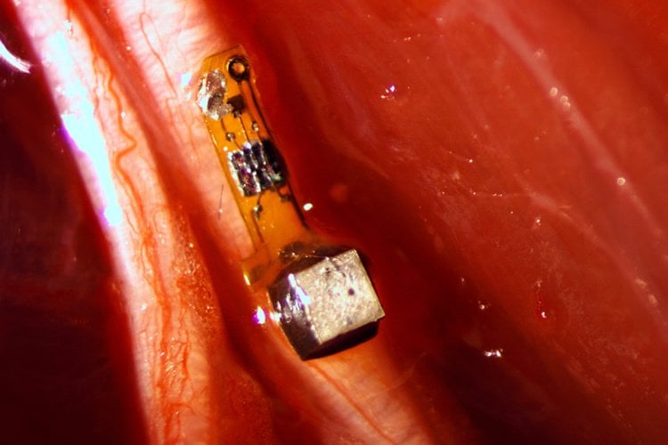
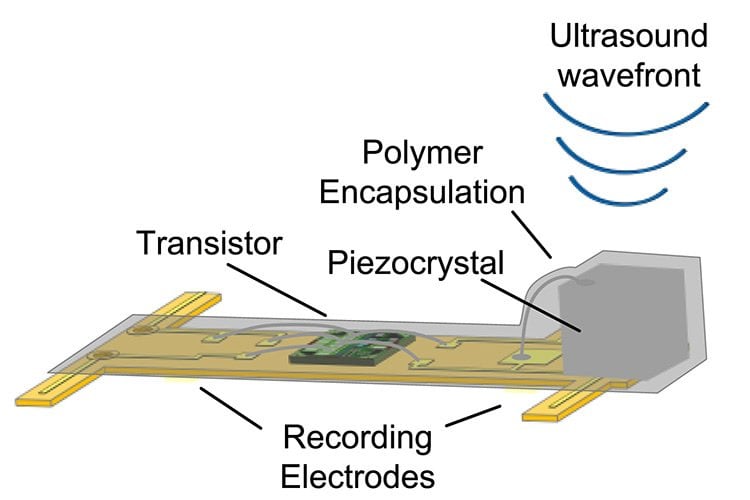
The sensors, which the researchers have already shrunk to a 1 millimeter cube – about the size of a large grain of sand – contain a piezoelectric crystal that converts ultrasound vibrations from outside the body into electricity to power a tiny, on-board transistor that is in contact with a nerve or muscle fiber. A voltage spike in the fiber alters the circuit and the vibration of the crystal, which changes the echo detected by the ultrasound receiver, typically the same device that generates the vibrations. The slight change, called backscatter, allows them to determine the voltage.
Motes sprinkled thoughout the body
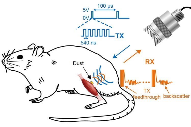
In their experiment, the UC Berkeley team powered up the passive sensors every 100 microseconds with six 540-nanosecond ultrasound pulses, which gave them a continual, real-time readout. They coated the first-generation motes – 3 millimeters long, 1 millimeter high and 4/5 millimeter thick – with surgical-grade epoxy, but they are currently building motes from biocompatible thin films which would potentially last in the body without degradation for a decade or more.
While the experiments so far have involved the peripheral nervous system and muscles, the neural dust motes could work equally well in the central nervous system and brain to control prosthetics, the researchers say. Today’s implantable electrodes degrade within 1 to 2 years, and all connect to wires that pass through holes in the skull. Wireless sensors – dozens to a hundred – could be sealed in, avoiding infection and unwanted movement of the electrodes.
“The original goal of the neural dust project was to imagine the next generation of brain-machine interfaces, and to make it a viable clinical technology,” said neuroscience graduate student Ryan Neely. “If a paraplegic wants to control a computer or a robotic arm, you would just implant this electrode in the brain and it would last essentially a lifetime.”
In a paper published online in 2013, the researchers estimated that they could shrink the sensors down to a cube 50 microns on a side – about 2 thousandths of an inch, or half the width of a human hair. At that size, the motes could nestle up to just a few nerve axons and continually record their electrical activity.
“The beauty is that now, the sensors are small enough to have a good application in the peripheral nervous system, for bladder control or appetite suppression, for example,“ Carmena said. “The technology is not really there yet to get to the 50-micron target size, which we would need for the brain and central nervous system. Once it’s clinically proven, however, neural dust will just replace wire electrodes. This time, once you close up the brain, you’re done.“
The team is working now to miniaturize the device further, find more biocompatible materials and improve the surface transceiver that sends and receives the ultrasounds, ideally using beam-steering technology to focus the sounds waves on individual motes. They are now building little backpacks for rats to hold the ultrasound transceiver that will record data from implanted motes.
They’re also working to expand the motes’ ability to detect non-electrical signals, such as oxygen or hormone levels.
“The vision is to implant these neural dust motes anywhere in the body, and have a patch over the implanted site send ultrasonic waves to wake up and receive necessary information from the motes for the desired therapy you want,” said Dongjin Seo, a graduate student in electrical engineering and computer sciences. “Eventually you would use multiple implants and one patch that would ping each implant individually, or all simultaneously.”
Ultrasound vs radio
Maharbiz and Carmena conceived of the idea of neural dust about five years ago, but attempts to power an implantable device and read out the data using radio waves were disappointing. Radio attenuates very quickly with distance in tissue, so communicating with devices deep in the body would be difficult without using potentially damaging high-intensity radiation.
Marharbiz hit on the idea of ultrasound, and in 2013 published a paper with Carmena, Seo and their colleagues describing how such a system might work. “Our first study demonstrated that the fundamental physics of ultrasound allowed for very, very small implants that could record and communicate neural data,” said Maharbiz. He and his students have now created that system.
University of California, Berkeley engineers have built the first dust-sized, wireless sensors that can be implanted in the body, bringing closer the day when a Fitbit-like device could monitor internal nerves, muscles or organs in real time.
“Ultrasound is much more efficient when you are targeting devices that are on the millimeter scale or smaller and that are embedded deep in the body,” Seo said. “You can get a lot of power into it and a lot more efficient transfer of energy and communication when using ultrasound as opposed to electromagnetic waves, which has been the go-to method for wirelessly transmitting power to miniature implants”
“Now that you have a reliable, minimally invasive neural pickup in your body, the technology could become the driver for a whole gamut of applications, things that today don’t even exist,“ Carmena said.
Other co-authors of the Neuron paper are graduate student Konlin Shen, undergraduate Utkarsh Singhal and UC Berkeley professors Elad Alon and Jan Rabaey.
Funding: The work was supported by the Defense Advanced Research Projects Agency of the Department of Defense.
Source: Robert Sanders – UC Berkeley
Image Source: This NeuroscienceNews.com images are adapted from the UC Berkeley press release and credited to Ryan Neely.
Video Source: The video is credited to UC Berkeley.
Original Research: Full open access research for “Wireless Recording in the Peripheral Nervous System with Ultrasonic Neural Dust” by Dongjin Seo, Ryan M. Neely, Konlin Shen, Utkarsh Singhal, Elad Alon, Jan M. Rabaey, Jose M. Carmena, and Michel M. Maharbiz in Neuron. Published online August 3 2016 doi:10.1016/j.neuron.2016.06.034
[cbtabs][cbtab title=”MLA”]UC Berkeley. “Sprinking of Neural Dust Opens Door to Electroceuticals.” NeuroscienceNews. NeuroscienceNews, 3 August 2016.
<https://neurosciencenews.com/neural-dust-wireless-sensors-4775/>.[/cbtab][cbtab title=”APA”]UC Berkeley. (2016, August 3). Sprinking of Neural Dust Opens Door to Electroceuticals. NeuroscienceNews. Retrieved August 3, 2016 from https://neurosciencenews.com/neural-dust-wireless-sensors-4775/[/cbtab][cbtab title=”Chicago”]UC Berkeley. “Sprinking of Neural Dust Opens Door to Electroceuticals.” https://neurosciencenews.com/neural-dust-wireless-sensors-4775/ (accessed August 3, 2016).[/cbtab][/cbtabs]
Abstract
Wireless Recording in the Peripheral Nervous System with Ultrasonic Neural Dust
Highlights
•First in vivo electrophysiological recordings with neural dust motes
•Passive, wireless, and battery-less EMG and ENG recording with mm-scale devices
•Recorded signals transmitted via ultrasonic backscatter from implanted neural dust motes
•Ultrasound as a scalable means of providing wireless power and communication
Summary
The emerging field of bioelectronic medicine seeks methods for deciphering and modulating electrophysiological activity in the body to attain therapeutic effects at target organs. Current approaches to interfacing with peripheral nerves and muscles rely heavily on wires, creating problems for chronic use, while emerging wireless approaches lack the size scalability necessary to interrogate small-diameter nerves. Furthermore, conventional electrode-based technologies lack the capability to record from nerves with high spatial resolution or to record independently from many discrete sites within a nerve bundle. Here, we demonstrate neural dust, a wireless and scalable ultrasonic backscatter system for powering and communicating with implanted bioelectronics. We show that ultrasound is effective at delivering power to mm-scale devices in tissue; likewise, passive, battery-less communication using backscatter enables high-fidelity transmission of electromyogram (EMG) and electroneurogram (ENG) signals from anesthetized rats. These results highlight the potential for an ultrasound-based neural interface system for advancing future bioelectronics-based therapies.
“Wireless Recording in the Peripheral Nervous System with Ultrasonic Neural Dust” by Dongjin Seo, Ryan M. Neely, Konlin Shen, Utkarsh Singhal, Elad Alon, Jan M. Rabaey, Jose M. Carmena, and Michel M. Maharbiz in Neuron. Published online August 3 2016 doi:10.1016/j.neuron.2016.06.034


