

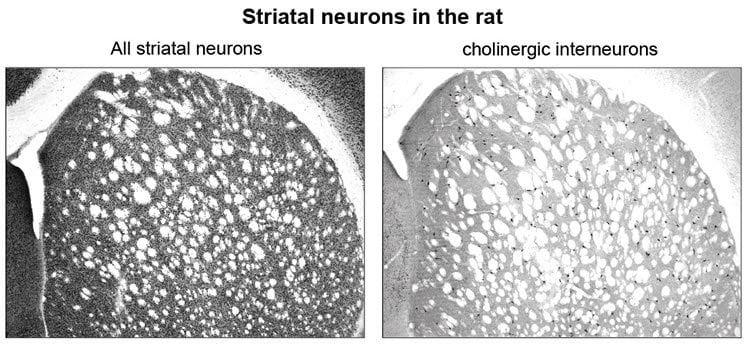
Behavioral flexibility, the ability to change strategy when the rules change, is controlled by specific neurons in the brain, Researchers at the Okinawa Institute of Science and Technology Graduate University (OIST) have confirmed. Cholinergic interneurons are rare – they make up just one to two percent of the neurons in the striatum, a key part of the brain involved with higher-level decision-making. Scientists have suspected they play a role in changing strategies, and researchers at OIST recently confirmed this with experiments. Their findings were published in the Journal of Neuroscience.
“Not much is known about these neurons,” said Sho Aoki, a post-doctoral researcher at OIST and lead author of the paper. “But we now have clear evidence that they play a key role in remaining flexible in this ever-changing world.”
Previous studies tried to identify the role of cholinergic interneurons by recording brain wave activity during behavioral tasks. While that can strongly indicate specific neurons are correlated with a particular behavior, it is not definitive. In this study, Aoki killed cholinergic interneurons with a toxin that directly targets them, and then observed how rats reacted to rule changes compared with normal rats with intact neurons. “Our experiments show direct causation, not correlation,” Aoki said.
Rats with and without damaged neurons were given tasks for several weeks – they had to press either lever A or B to get a sugar pellet reward. During the first few days, Lever A always resulted in a reward. Both groups of rats had no problem learning the initial strategy to get the sugar pellet – press Lever A.
But then, the rules of the game changed. A novel stimulus was introduced – a light flashed above the correct lever, which oscillated between Lever A and B (Fig. 2A). To get their sugar fix, the rats had to shift strategy and pay attention to this new information. While normal rats quickly responded to the light, rats with damaged neurons could not. The latter group continued to repeat the strategy they had already learned, and were disinclined to explore what the light meant.
In another test, a light cue that had been flashing in a meaningless pattern during the initial learning phase switched to signaling the correct lever to push for reward (Fig. 2B). This meant to maximize rewards, and the animals should now pay attention to a stimulus they previously ignored. Again, the control rats had no problem adapting to this rule change, but the damaged rats stuck to their original strategy, even though it meant fewer rewards. They also decreased exploring what might increase their chance of success.
Interestingly, rats with neurons damaged in the dorsomedial part of the striatum had greater difficulty paying attention to previously irrelevant light cues (Fig. 2B). Rats with neurons damaged in the ventral part of the striatum had a harder time reacting to novel stimulants (Fig. 2A).

“This indicates that cholinergic interneurons throughout the striatum play a common role, namely inhibiting old rules and encouraging exploration, but different regions of the striatum are activated depending on the situation and type of stimulus,” Aoki said.
The research findings might help researchers and medical professionals who investigate aging. “Since cholinergic interneurons degenerate with age, this work may provide a clue for understanding the decline in mental flexibility that occurs with advancing age,” said Prof. Jeff Wickens, head of OIST’s Neurobiology Research Unit and senior paper author.
Source: Laura Petersen – OIST
Image Credit: Images are credited to Laura Petersen/OIST
Original Research: Abstract for “Role of Striatal Cholinergic Interneurons in Set-Shifting in the Rat” by Sho Aoki, Andrew W. Liu, Aya Zucca, Stefano Zucca, and Jeffery R. Wickens in Journal of Neuroscience. Published online June 24 2015 doi:10.1523/JNEUROSCI.0490-15.2015
Abstract
Role of Striatal Cholinergic Interneurons in Set-Shifting in the Rat
The ability to change strategies in different contexts is a form of behavioral flexibility that is crucial for adaptive behavior. The striatum has been shown to contribute to certain forms of behavioral flexibility such as reversal learning. Here we report on the contribution of striatal cholinergic interneurons—a key element in the striatal neuronal circuit—to strategy set-shifting in which an attentional shift from one stimulus dimension to another is required. We made lesions of rat cholinergic interneurons in dorsomedial or ventral striatum using a specific immunotoxin and investigated the effects on set-shifting paradigms and on reversal learning. In shifting to a set that required attention to a previously irrelevant cue, lesions of dorsomedial striatum significantly increased the number of perseverative errors. In this condition, the number of never-reinforced errors was significantly decreased in both types of lesions. When shifting to a set that required attention to a novel cue, rats with ventral striatum lesions made more perseverative errors. Neither lesion impaired learning of the initial response strategy nor a subsequent switch to a new strategy when response choice was indicated by a previously relevant cue. Reversal learning was not affected. These results suggest that in set-shifting the striatal cholinergic interneurons play a fundamental role, which is dissociable between dorsomedial and ventral striatum depending on behavioral context. We propose a common mechanism in which cholinergic interneurons inhibit neurons representing the old strategy and enhance plasticity underlying exploration of a new rule.
“Role of Striatal Cholinergic Interneurons in Set-Shifting in the Rat” by Sho Aoki, Andrew W. Liu, Aya Zucca, Stefano Zucca, and Jeffery R. Wickens in Journal of Neuroscience. Published online June 24 2015 doi:10.1523/JNEUROSCI.0490-15.2015


