Summary: Researchers have discovered how the brain controls our breathing in response to changing oxygen and carbon dioxide levels in the blood.
Source: eLife.
Scientists from Karolinska Institutet, Sweden, have discovered how the brain controls our breathing in response to changing oxygen and carbon dioxide levels in the blood.
The control of breathing is essential for life. Without an adequate response to increased carbon dioxide levels, people can suffer from breathing disturbances, sickness, and panic. In worst-case scenarios, it can lead to premature death, as in sudden infant death syndrome.
There has been some debate over how the brain controls breathing. Now, a new study in mice, to be published in the journal eLife, shows that when exposed to decreased oxygen or increased carbon dioxide levels, the brain releases a small molecule called Prostaglandin E2 (PGE2) to help protect itself and regulate breathing.
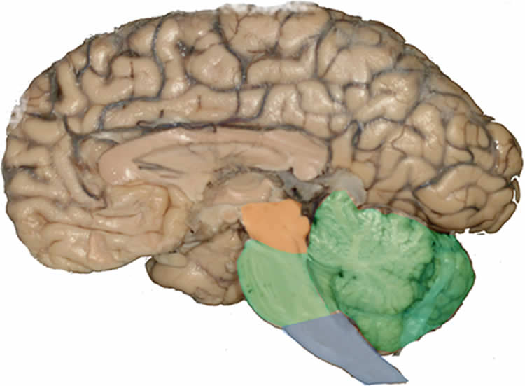
To discover this mechanism, the researchers grew a section of a mouse’s brainstem (the central trunk of the brain) in a type of dish. The slice contained an arrangement of nerve and supporting cells that allowed it to ‘breathe’ for three weeks. During this time, the team monitored the cells and their behaviour in response to changes in the environment.
“Our novel brainstem culture first revealed that cells responsible for breathing operate in a small-world network. Groups of these cells work very closely with each other, with each group interconnected by a few additional cells that appear to work as hubs. This networking activity and the rhythmic respiratory motor output it generated were preserved for the full three weeks, suggesting that our brainstem can be used for long-term studies of respiratory neural network activity,” explains David Forsberg, PhD student and first author of the study.
“Secondly, we saw that exposure to different substances made the brainstem breathe faster or slower. Perhaps most interesting was its response to carbon dioxide, which triggered a release of PGE2. Here, PGE2 acted as a signaling molecule that increased breathing activity in the carbon dioxide-sensitive brainstem region, leading to slower and deeper breaths, or ‘sighs’.”
These new insights have important implications for babies, who experience significantly reduced levels of oxygen during birth. At this stage, PGE2 protects the brain and prepares the brainstem to generate deep sigh-like breath intakes, resulting in the first breaths of air following birth.
The study also reveals a novel pathway linking the inflammatory and respiratory systems. PGE2 is released during inflammation and fever, which can dysregulate breathing patterns and interfere with normal responses to carbon dioxide. This can in turn cause disturbed and even dangerous halts in breathing.
“Our findings go some way to explaining how and why our breathing responses to imbalanced oxygen and carbon dioxide levels are impaired during infectious episodes. It also helps further our understanding of why infection can inhibit breathing so severely in new-born babies,” says Eric Herlenius, Professor at Karolinska Institutet’s Department of Women’s and Children’s Health, and senior author of the paper.
“We now want to find out how breaths form and develop during episodes of inflammation. This could be useful for researching potential new ways to save babies’ lives when they are unable to catch their breaths.”
Funding: Karolinska Institutet, Vetenskapsrådet, VINNOVA, Stockholms Läns Landsting, Hjärnfonden, The Swedish National Heart and Lung Foundation, Knut och Alice Wallenbergs Stiftelse funded this study.
Source: Emily Packer – eLife
Image Source: This NeuroscienceNews.com image is credited to Grinberg LT, Rueb U and Heinsen H and is licensed CC BY 3.0.
Original Research: The findings will be presented at 10th FENS Forum of Neuroscience 2016 in Copenhagen, Denmark between July 2 – 6, 2016.
Abstract for “CO2-evoked release of PGE2 modulates sighs and inspiration as demonstrated in brainstem organotypic culture” by David Forsberg, Zachi Horn, Evangelia Tserga, Erik Smedler, Gilad Silberberg, Yuri Shvarev, Kai Kaila, Per Uhlén and Eric Herlenius in eLife. Published online July 5 2016 doi:10.7554/eLife.14170
[cbtabs][cbtab title=”MLA”]eLife. “New Brainstem Model Reveals How the Brain Controls Breathing.” NeuroscienceNews. NeuroscienceNews, 5 July 2016.
<https://neurosciencenews.com/brainstem-model-breathing-4620/>.[/cbtab][cbtab title=”APA”]eLife. (2016, July 5). New Brainstem Model Reveals How the Brain Controls Breathing. NeuroscienceNew. Retrieved July 5, 2016 from https://neurosciencenews.com/brainstem-model-breathing-4620/[/cbtab][cbtab title=”Chicago”]eLife. “New Brainstem Model Reveals How the Brain Controls Breathing.” https://neurosciencenews.com/brainstem-model-breathing-4620/ (accessed July 5, 2016).[/cbtab][/cbtabs]
Abstract
CO2-evoked release of PGE2 modulates sighs and inspiration as demonstrated in brainstem organotypic culture
Inflammation-induced release of prostaglandin E2 (PGE2) changes breathing patterns and the response to CO2 levels. This may have fatal consequences in newborn babies and result in sudden infant death. To elucidate the underlying mechanisms, we present a novel breathing brainstem organotypic culture that generates rhythmic neural network and motor activity for 3 weeks. We show that increased CO2 elicits a gap junction-dependent release of PGE2. This alters neural network activity in the preBötzinger rhythm-generating complex and in the chemosensitive brainstem respiratory regions, thereby increasing sigh frequency and the depth of inspiration. We used mice lacking eicosanoid prostanoid 3 receptors (EP3R), breathing brainstem organotypic slices and optogenetic inhibition of EP3R+/+cells to demonstrate that the EP3R is important for the ventilatory response to hypercapnia. Our study identifies a novel pathway linking the inflammatory and respiratory systems, with implications for inspiration and sighs throughout life, and the ability to autoresuscitate when breathing fails.
“CO2-evoked release of PGE2 modulates sighs and inspiration as demonstrated in brainstem organotypic culture” by David Forsberg, Zachi Horn, Evangelia Tserga, Erik Smedler, Gilad Silberberg, Yuri Shvarev, Kai Kaila, Per Uhlén and Eric Herlenius in eLife. Published online July 5 2016 doi:10.7554/eLife.14170