Neuroscientists know that some connections in the brain are pruned through neural development. Function gives rise to structure, according to the textbooks. But scientists at the Virginia Tech Carilion Research Institute have discovered that the textbooks might be wrong.
Their results were published today in Cell Reports.
“Retinal neurons associated with vision generate connections in the brain, and as the brain develops it strengthens and maintains some of those connections more than others. The disused connections are eliminated,” said Michael Fox, an associate professor at the Virginia Tech Carilion Research Institute who led the study. “We found that this activity-dependent pruning might not be as simple as we’d like to believe.”
Fox and his team of researchers used two different techniques to examine how retinal ganglion cells – neurons that live in the retina and transmit visual information to the visual centers in the brain – develop in a mouse model.
“It’s widely accepted that synaptic connections from about 20 retinal ganglion cells converge onto cells in the lateral geniculate nucleus during development, but that number reduces to just one or two by the third week of a mouse’s life,” Fox said. “It was thought that the mature retinal ganglion cells develop several synaptic terminals that cluster around information exchange points.”
The theory of several terminals blossoming from the same retinal ganglion cell had not been proved, though, so Fox and his researchers decided to follow the terminals to their roots.
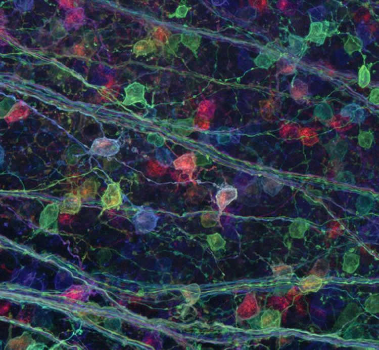
Using a technique dubbed “brainbow,” the scientists tagged the terminals with proteins that fluoresce different colors. The researchers thought one color, representing the single source of the many terminals, would dominate in the clusters. Instead, several different colors appeared together, intertwined but distinct.
“The samples showed a true ‘brainbow,'” said Aboozar Monavarfeshani, a graduate student in Fox’s laboratory who tagged the terminals. “I could see, right in front of me, something very different than the concept I learned from my textbooks.”
The results showed individual terminals from more than one retinal ganglion cell in a mature mouse brain.
The study is a direct contradiction to some other research indicating neural development weeds out most connections between retinal ganglion axons and target cells in the brain, and Fox and his team have more questions.
“Is this a discrepancy a technical issue with the different types of approaches applied in all of these disparate studies?” Fox asked. “Possibly, but perhaps it’s more likely that retinal ganglion cells are more complex than previously thought.”
Along with the brainbow technique, Fox’s team also imaged these synaptic connections with electron microscopy.
Sarah Hammer, currently a sophomore at Virginia Tech, traced individual retinal terminals through hundreds of serial images.
The data confirmed the results from “brainbow” analysis – retinal axons from numerous retinal ganglion cells remained present on adult brain cells.
“These results are not what we expected, and they will force us to reevaluate our understanding of the architecture and flow of visual information through neural pathways,” Fox said. “The dichotomy of these results also sheds important light on the benefits of combining approaches to understand complicated problems in science.”
Albert Pan, an assistant professor in the Medical College of Georgia at Georgia Regents University, who is an expert in neural circuitry development, said the results are unexpected.
“The research provides strong evidence for multiple innervation and calls for a reevaluation of the current understanding of information flow and neural circuit maturation in the visual system” said Pan, who was not involved in the study. “The paper probably generates more questions than it answers, which is a hallmark of an exciting research study.”
The research continues, as Fox’s team works to understand exactly how many retinal terminals converge and how they might convey information differently. Once the scientists understand the intricacies of the brain’s visual circuitry, they might be able to start developing therapeutics for when it goes wrong.
“The lesson in this particular study is that no single technique gives us all the right answers,” Fox said. “Science is never as simple as we like to make it seem.”
Source: Paula Brewer Byron – Virginia Tech
Image Source: The image is credited to Virginia Tech
Original Research: Full open access research for “Multiple Retinal Axons Converge onto Relay Cells in the Adult Mouse Thalamus” by Sarah Hammer, Aboozar Monavarfeshani, Tyler Lemon, Jianmin Su, and Michael Andrew Fox in Cell Reports. Published online August 27 2015 doi:10.1016/j.celrep.2015.08.003
Abstract
Multiple Retinal Axons Converge onto Relay Cells in the Adult Mouse Thalamus
Highlights
•Brainbow-based technology can be used to differentially label retinal terminals
•Clusters of retinal terminals originate from numerous uniquely labeled RGCs
•Complex retinogeniculate synapses contain terminals from >10 distinct retinal axons
•A higher level of convergence exists on relay cells than previously appreciated
Summary
Activity-dependent refinement of neural circuits is a fundamental principle of neural development. This process has been well studied at retinogeniculate synapses—synapses that form between retinal ganglion cells (RGCs) and relay cells within the dorsal lateral geniculate nucleus. Physiological studies suggest that shortly after birth, inputs from ∼20 RGCs converge onto relay cells. Subsequently, all but just one to two of these inputs are eliminated. Despite widespread acceptance, this notion is at odds with ultrastructural studies showing numerous retinal terminals clustering onto relay cell dendrites in the adult. Here, we explored this discrepancy using brainbow AAVs and serial block face scanning electron microscopy (SBFSEM). Results with both approaches demonstrate that terminals from numerous RGCs cluster onto relay cell dendrites, challenging the notion that only one to two RGCs innervate each relay cell. These findings force us to re-evaluate our understanding of subcortical visual circuitry.
“Multiple Retinal Axons Converge onto Relay Cells in the Adult Mouse Thalamus” by Sarah Hammer, Aboozar Monavarfeshani, Tyler Lemon, Jianmin Su, and Michael Andrew Fox in Cell Reports. Published online August 27 2015 doi:10.1016/j.celrep.2015.08.003