

How does the brain make connections, and how does it maintain them? Cambridge neuroscientists and mathematicians are using a variety of techniques to understand how the brain ‘wires up’, and what it might be able to tell us about degeneration in later life.
To read these words, light is first refracted by the cornea, through the pupil in the iris and onto the lens, which focuses images onto the retina. The images are received by light-sensitive cells in the retina, which transmit impulses to the brain. These impulses are carried by a set of neurons called the retinal ganglion cells. Once the impulses reach the brain, the brain then has to piece together the information it receives into an understandable image. All of this happens in a fraction of a second.
Information travels from the retina to the brain via axons – the long, threadlike parts of neurons – sent out by the retinal ganglion cells. During embryonic development, axons are sent out to find their specific targets in the brain, so that images can be processed.
For an axon in a growing embryo, the journey from retina to brain is not a straightforward one. It’s a very long way for a tiny axon, through a constantly changing series of environments that it has never encountered before. So how do axons know where to go, and what can it tell us about how the brain is made and maintained?
Two University of Cambridge researchers, Professor Christine Holt of the Department of Physiology, Development and Neuroscience, and Dr Stephen Eglen of the Department of Applied Mathematics and Theoretical Physics, are taking two different, but complementary, approaches to these questions.
With funding from the European Research Council and the Wellcome Trust, Holt’s research group is aiming to better understand the molecular and cellular mechanisms that guide and maintain axon growth, which in turn will aid better understanding of how nerve connections are first established. “It’s an impressive navigational feat,” says Holt. “The pathway between the retina and the brain may look homogeneous, but in reality it’s like a patchwork quilt of different molecular domains.”
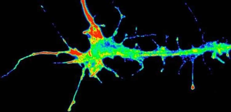
On the pathway through this patchwork quilt, there is a set of distinct beacons, breaking the axon’s journey down into separate steps. Every time the growing axon reaches a new beacon, it has to make a decision about which way to go. At the tip of the axon is a growth cone, which ‘sniffs out’ certain chemical signals emitted from the beacons, helping it to steer in the right direction.
The growth cones are receptive to certain signals and blind to others, so depending on what the axon encounters when it reaches a particular beacon, it will behave in a certain way. Holt’s research group uses a variety of techniques to determine what the signals are at the steering points where axons alter their direction of growth or their behaviour, such as the optic chiasm where certain axons cross to the opposite side of the brain, or at the point where they first leave the eye.
While Holt uses experiments to understand the development of the visual system, Eglen uses mathematical models as a complementary technique to try to answer the same questions. “You’ve got much more freedom in a theoretical model than you do in an experiment,” he says. “A common experimental approach is to remove something genetically and see what happens. I think of that a little like taking the battery out of your car. Doing that will tell you that the battery is necessary for the car to function, but it doesn’t really tell you why.”
Theoretical models allow researchers to approach the questions around neural development from a different angle. To capture the essence of the neural system, they try to represent the building blocks of development and see what kind of behaviour would result.
But no model yet can fully capture the complexities of how the visual system develops, which Eglen views not only as a challenge for him as a mathematician, but also as a challenge back to the experimental community. “It had been thought that if we built a model and took out all of the guidance molecules, there would be no topographic order whatsoever,” says Eglen. “But instead we found that there is still residual order in how the neurons are wired up, so there must be extra molecules or mechanisms that we don’t know about. What we’re trying to do is to take biology and put it into computers so that we can really test it.”
“In the past 15–20 years, there’s been a revolution in terms of being able to identify the specific molecules that act as guidance receptors or signals, but there’s still so much we don’t yet know, which is why we’re using both theoretical and experimental techniques to answer these questions,” says Holt. “And in addition to this question of wiring, we’re also looking at the problem of mapping – how do the terminal ends of the axons find their ultimate destination in the brain?”
Holt’s group has found that the same guidance molecule can have different roles depending on what aspect of growth is going on – but the question then becomes how do you wire the brain with so few molecules?
Adding to the complexity was another puzzling discovery – that the growth cones of axons can make proteins. Previous knowledge held that new proteins could be synthesised only within the main cellular part of each neuron, the cell body (where the nucleus is located), and then transported into axons. However, Holt’s group found that the growth cones of axons are also capable of synthesising proteins ‘on demand’ when they encounter new guidance beacons, suggesting that messenger RNA (mRNA) molecules play a role in helping axons to navigate to their correct destinations. mRNAs are the molecules from which new proteins are synthesised, and further experiments found that axons contain hundreds or even thousands of different types of this nuclear material.
In addition to their role in axon growth when the brain is wiring itself up during development, certain types of mRNA are also important in maintaining the connections in the adult brain, by keeping mitochondria – the energy-producing ‘batteries’ of cells – healthy, which, in turn, keeps axons healthy.
“It is a whole new view to the idea of degeneration in later life – a lot of different components have to work together to get local protein synthesis to work, so if just one of those components fails, degeneration can occur,” says Holt. “We’ve also found that many of the types of mRNA that are being translated in axons are the same ones that you see in diseases like Huntingdon’s and Parkinson’s, so basic knowledge of this sort is essential for the development of clinical therapies in nerve repair and for understanding these and other neurodegenerative disorders.”
Source: Sarah Collins – University of Cambridge
Image Credit: The image is credited to K-M. Leung.


