

Human-specific network may have evolved to strengthen social communication.
Researchers at Columbia University Medical Center (CUMC) have found that key parts of the human brain network that give us the power to control and redirect our attention—a core cognitive ability—may be unique to humans. The research, which was published in the July 13 online edition of the Proceedings of the National Academy of Sciences, suggests that the network may have evolved in response to increasingly complex social cues.
“The human brain is powerful, but even it cannot make sense of the entire sum of stimuli that bombard our senses,” said Vincent P. Ferrera, PhD, the study’s senior author. Dr. Ferrera is a principal investigator at Columbia’s Mortimer B. Zuckerman Mind Bran Behavior Institute and associate professor in the department of neuroscience (in psychiatry) at CUMC. “Instead, it selects and prioritizes information based on what is needed at any given moment—this is called attention. And while attention is a fundamental characteristic of human cognition, and something that we use all the time, the underlying brain circuits that give us this ability remain largely unclear.”
In order to better understand these circuits, the authors compared the brains of primate and human subjects during a specific attention-seeking task. In so doing, they uncovered key clues about these so-called ‘attention networks’: how they evolved and how they underlie human cognitive abilities—and are already using this information to test what role they may play in psychiatric disorders.
In the human brain, there are two main attention networks: the dorsal attention network (DAN) and ventral attention network (VAN). “The DAN is in charge of directing your attention to something specific, and when it’s active, the VAN is silent,” explained Gaurav Patel, MD/PhD, a postdoctoral fellow in the Department of Psychiatry and the study’s first author. “This keeps you focused and limits your distraction. But if you see something that is new, unique, or behaviorally relevant, the VAN will switch on. This give-and-take between the DAN and VAN allows us to reorient our attention to what is most important.”
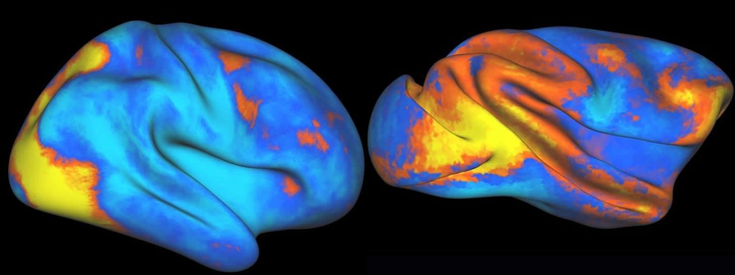
In a series of experiments, first using a primate model at Washington University in St. Louis, and later with human subjects at CUMC, the researchers used functional magnetic resonance imaging (fMRI) to map brain activity. Before the start of a session, the subject memorized a specific target object. During the session, a stream of images appeared on various parts of a screen. The subject pushed a button when the target object appeared on the screen. fMRI technology allowed the researchers to see when various parts of the brain ‘switched on’ while the subject performed the task. And the results were surprising.
“The fMRI showed striking differences between the two species, which was wholly unexpected,” said Dr. Patel. “We were giving both the human and primate subjects the exact same task—their brain activity should have been more similar. That’s when we realized there was something else going on.”
First, they realized that the VAN—located in the right hemisphere of the human brain—had no equivalent in the primate brain. They also noticed the DAN had expanded in the human subjects, and observed enhanced cross talk between the human subjects’ brain hemispheres.
“Taken together, these findings suggests that at some point in our evolutionary history, we evolved an additional attention network—perhaps in order to better process the world around us,” said Dr. Patel.
The human social world is far more complex than any of our primate relatives. It is possible this network evolved to help us understand increasingly complex social cues, such as the subtle twitch of an eyebrow, or a shift in gaze, say the researchers.

Armed with this new information, Dr. Patel is now examining the attention networks of people with schizophrenia—a disorder characterized in part by poor social function.
“Persons with schizophrenia have difficulty expressing emotions, responding to social cues and often become socially isolated,” said Dr. Patel. “Right now we’re testing whether these individuals have an impairment to how the VAN is organized or communicates with the rest of the brain. Because if they do, we could potentially tailor a treatment strategy to mitigate that impairment, and maybe even restore some level of social communication that the patient lacked.”
Funding:This research was supported by the Levy Foundation, the American Psychiatric Foundation, the National Institute of Mental Health (MH086466-04, MH018870-25, MH102471, MH096482) and the National Eye Institute (EY012135).
The authors report no financial or other conflicts of interest.
Source: Anne Holden, PhD – Columbia University Medical Center
Image Credit: The images are credited to Gaurav Patel/New York State Psychiatric Institute/Columbia University Medical Center
Original Research: Abstract for “Functional evolution of new and expanded attention networks in humans” by Gaurav H. Patel, Danica Yang, Emery C. Jamerson, Lawrence H. Snyder, Maurizio Corbetta, and Vincent P. Ferrera in PNAS. Published online July 13 2015 doi:10.1073/pnas.1420395112
Abstract
Functional evolution of new and expanded attention networks in humans
Macaques are often used as a model system for invasive investigations of the neural substrates of cognition. However, 25 million years of evolution separate humans and macaques from their last common ancestor, and this has likely substantially impacted the function of the cortical networks underlying cognitive processes, such as attention. We examined the homology of frontoparietal networks underlying attention by comparing functional MRI data from macaques and humans performing the same visual search task. Although there are broad similarities, we found fundamental differences between the species. First, humans have more dorsal attention network areas than macaques, indicating that in the course of evolution the human attention system has expanded compared with macaques. Second, potentially homologous areas in the dorsal attention network have markedly different biases toward representing the contralateral hemifield, indicating that the underlying neural architecture of these areas may differ in the most basic of properties, such as receptive field distribution. Third, despite clear evidence of the temporoparietal junction node of the ventral attention network in humans as elicited by this visual search task, we did not find functional evidence of a temporoparietal junction in macaques. None of these differences were the result of differences in training, experimental power, or anatomical variability between the two species. The results of this study indicate that macaque data should be applied to human models of cognition cautiously, and demonstrate how evolution may shape cortical networks.
“Functional evolution of new and expanded attention networks in humans” by Gaurav H. Patel, Danica Yang, Emery C. Jamerson, Lawrence H. Snyder, Maurizio Corbetta, and Vincent P. Ferrera in PNAS. Published online July 13 2015 doi:10.1073/pnas.1420395112


