

Summary: According to researchers, if the ventral tegmental area is unable to produce the correct amount of dopamine for the hippocampus, it will not work efficiently and the impairment can result in dementia.
Source: University of Sheffield.
A new link between diminished input from dopamine-firing cells deep inside the brain and the ability to form new memories could be crucial in detecting the earliest signs of Alzheimer’s disease.
Scientists from the National Institute for Health Research (NIHR) Sheffield Biomedical Research Centre at the University of Sheffield have discovered a loss of cells that use dopamine – a neurotransmitter that has a number of functions including regulating movement and emotional responses – may cause the part of the brain responsible for forming new memories to function less effectively.
The findings could revolutionise screening for the early signs of Alzheimer’s disease – which affects more than 520,000 people in the UK – changing the way brain scans are acquired and interpreted as well as using different memory tests.
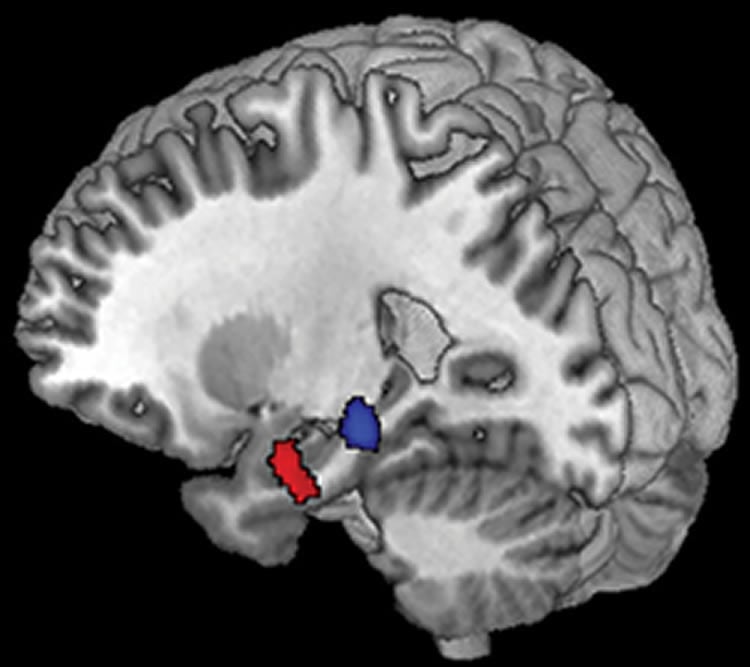
Lead author of the study, Professor Annalena Venneri, from the Sheffield Institute for Translational Neuroscience (SITraN) at the University of Sheffield, said: “Our findings suggest that if a small area of brain cells, called the ventral tegmental area, does not produce the right amount of dopamine for the hippocampus, a small organ located within the brain’s temporal lobe, it will not work efficiently.
“The hippocampus is associated with forming new memories, therefore these findings are crucial to the early detection of Alzheimer’s disease. The results point at a change which happens very early on, which might trigger Alzheimer’s disease.
“This is the first study to demonstrate such a link in humans.”
Professor Venneri and fellow lead author Dr Matteo De Marco acquired 3Tesla Magnetic Resonance Imaging (MRI) scans on 51 healthy adults, 30 patients with a diagnosis of mild cognitive impairment, and 29 patients with a diagnosis of Alzheimer’s disease. 3Tesla MRIs are twice the normal strength of normal MRI scans generating the highest quality images.
The results showed a key link between the size and function of the ventral tegmental area, the size of the hippocampus and the ability to learn new material.
“More studies are necessary, but these findings could potentially lead to a new way of screening the elderly population for early signs of Alzheimer’s disease, changing the way brain scans are acquired and interpreted and using different memory tests,” said Professor Venneri who is also an Honorary Consultant at Sheffield Teaching Hospital NHS Foundation Trust.
“Another possible benefit is that it might lead to a different treatment option with the potential to change or halt the course of the disease very early, before major symptoms manifest.
“We now want to establish how early alterations in the ventral tegmental area can be seen and also test whether these alterations can be counteracted with treatments already available.”
Funding: This study has been completed at the NIHR Biomedical Research Centre (BRC) in Sheffield which is dedicated to translational neuroscience for chronic neurological disorders. The Centre was launched in April 2017.
The NIHR Sheffield Biomedical Research Centre is a research partnership between the University of Sheffield and Sheffield Teaching Hospitals NHS Foundation Trust, dedicated to improving the treatment and care of people living with chronic neurological disorders.
Source: University of Sheffield
Publisher: Organized by NeuroscienceNews.com.
Image Source: NeuroscienceNews.com image is credited to University of Sheffield.
Original Research: Abstract for “Volume and Connectivity of the Ventral Tegmental Area are Linked to Neurocognitive Signatures of Alzheimer’s Disease in Humans” by De Marco, Matteo; and Venneri, Annalena in Journal of Alzheimer’s Disease. Published online March 23 2018.
doi:10.3233/JAD-171018
[cbtabs][cbtab title=”MLA”]University of Sheffield “Detecting Diminishing Dopamine Firing Cells in Brain Could Reveal Earliest Signs of Alzheimer’s.” NeuroscienceNews. NeuroscienceNews, 27 March 2018.
<https://neurosciencenews.com/alzheimers-dopamine-neuron-8698/>.[/cbtab][cbtab title=”APA”]University of Sheffield (2018, March 27). Detecting Diminishing Dopamine Firing Cells in Brain Could Reveal Earliest Signs of Alzheimer’s. NeuroscienceNews. Retrieved March 27, 2018 from https://neurosciencenews.com/alzheimers-dopamine-neuron-8698/[/cbtab][cbtab title=”Chicago”]University of Sheffield “Detecting Diminishing Dopamine Firing Cells in Brain Could Reveal Earliest Signs of Alzheimer’s.” https://neurosciencenews.com/alzheimers-dopamine-neuron-8698/ (accessed March 27, 2018).[/cbtab][/cbtabs]
Abstract
Volume and Connectivity of the Ventral Tegmental Area are Linked to Neurocognitive Signatures of Alzheimer’s Disease in Humans
Background:There is an urgent need to identify the earliest biological changes within the neuropathological cascade of Alzheimer’s disease (AD) processes. Recent findings in a murine model of AD showed significant preclinical loss of dopaminergic neurons in the ventral tegmental area (VTA), accompanied by reduced hippocampal innervation and declining memory. It is unknown if these observations can be translated in humans. Objective:We tested the hypothesis that VTA volume is associated with the typical clinical markers of AD in a cohort of patients and healthy controls.
Methods:Structural and resting state functional MRI scans, and neuropsychological scores were acquired for 51 healthy adults, 30 patients with a diagnosis of mild cognitive impairment, and 29 patients with a diagnosis of AD dementia. VTA volume was quantified together with other control nuclei. The association between nuclei volume, hippocampal size, memory performance, and linguistic-executive skills was tested. The effect of VTA functional connectivity was also tested.
Results:VTA size, but not of control nuclei, yielded a strong association with both hippocampal size and memory competence (but not linguistic-executive performance), and this was particularly strong in healthy adults. In addition, functional connectivity between the VTA and hippocampus was significantly associated with both markers of AD.
Conclusion:Diminished dopaminergic VTA activity may be crucial for the earliest pathological features of AD and might suggest new strategies for early treatment. Memory encoding processes may represent cognitive operations susceptible to VTA neurodegeneration.


