


TAU researchers find unresponsive patients’ brains may recognize photographs of their family and friends.
Patients in a vegetative state are awake, breathe on their own, and seem to go in and out of sleep. But they do not respond to what is happening around them and exhibit no signs of conscious awareness. With communication impossible, friends and family are left wondering if the patients even know they are there.
Now, using functional magnetic resonance imaging (fMRI), Dr. Haggai Sharon and Dr. Yotam Pasternak of Tel Aviv University’s Functional Brain Center and Sackler Faculty of Medicine and the Tel Aviv Sourasky Medical Center have shown that the brains of patients in a vegetative state emotionally react to photographs of people they know personally as though they recognize them.
“We showed that patients in a vegetative state can react differently to different stimuli in the environment depending on their emotional value,” said Dr. Sharon. “It’s not a generic thing; it’s personal and autobiographical. We engaged the person, the individual, inside the patient.”
The findings, published in PLOS ONE, deepen our understanding of the vegetative state and may offer hope for better care and the development of novel treatments. Researchers from TAU’s School of Psychological Sciences, Department of Neurology, and Sagol School of Neuroscience and the Loewenstein Hospital in Ranaana contributed to the research.
Talking to the brain
For many years, patients in a vegetative state were believed to have no awareness of self or environment. But in recent years, doctors have made use of fMRI to examine brain activity in such patients. They have found that some patients in a vegetative state can perform complex cognitive tasks on command, like imagining a physical activity such as playing tennis, or, in one case, even answering yes-or-no questions. But these cases are rare and don’t provide any indication as to whether patients are having personal emotional experiences in such a state.
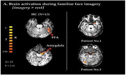
To gain insight into “what it feels like to be in a vegetative state,” the researchers worked with four patients in a persistent (defined as “month-long”) or permanent (persisting for more than three months) vegetative state. They showed them photographs of people they did and did not personally know, then gauged the patients’ reactions using fMRI, which measures blood flow in the brain to detect areas of neurological activity in real time. In response to all the photographs, a region specific to facial recognition was activated in the patients’ brains, indicating that their brains had correctly identified that they were looking at faces.
But in response to the photographs of close family members and friends, brain regions involved in emotional significance and autobiographical information were also activated in the patients’ brains. In other words, the patients reacted with activations of brain centers involved in processing emotion, as though they knew the people in the photographs. The results suggest patients in a vegetative state can register and categorize complex visual information and connect it to memories – a groundbreaking finding.
The ghost in the machine
However, the researchers could not be sure if the patients were conscious of their emotions or just reacting spontaneously. So they then verbally asked the patients to imagine their parents’ faces. Surprisingly, one patient, a 60-year-old kindergarten teacher who was hit by a car while crossing the street, exhibited complex brain activity in the face- and emotion-specific brain regions, identical to brain activity seen in healthy people. The researchers say her response is the strongest evidence yet that vegetative-state patients can be “emotionally aware.” A second patient, a 23-year-old woman, exhibited activity just in the emotion-specific brain regions. (Significantly, both patients woke up within two months of the tests. They did not remember being in a vegetative state.)
“This experiment, a first of its kind, demonstrates that some vegetative patients may not only possess emotional awareness of the environment but also experience emotional awareness driven by internal processes, such as images,” said Dr. Sharon.
Research focused on the “emotional awareness” of patients in a vegetative state is only a few years old. The researchers hope their work will eventually contribute to improved care and treatment. They have also begun working with patients in a minimally conscious state to better understand how regions of the brain interact in response to familiar cues. Emotions, they say, could help unlock the secrets of consciousness.
Notes about this neurology research
Contact: George Hunka – American Friends of Tel Aviv University
Source: American Friends of Tel Aviv University press release
Image Source: Image credited to Haggai Sharon, Yotam Pasternak, Eti Ben Simon, Michal Gruberger, Nir Giladi, Ben Zion Krimchanski, David Hassin, and Talma Hendler/PLOS ONE and adapted from the research paper.
Original Research: Full open access research for “Emotional Processing of Personally Familiar Faces in the Vegetative State” by Haggai Sharon, Yotam Pasternak, Eti Ben Simon, Michal Gruberger, Nir Giladi, Ben Zion Krimchanski, David Hassin, and Talma Hendler in PLOS ONE. Published online September 25 2013 doi:10.1371/journal.pone.0074711


