Summary: Researchers have identified a white matter pathway associated with steroacuity.
Source: NICT.
Researchers in the Center for Information and Neural Networks (CiNet), the National Institute of Information and Communications Technology (NICT, President: Hideyuki Tokuda, Ph.D.), and Osaka University (President: Shojiro Nishio, Ph.D.) have identified a human white matter pathway associated with individual variability in human stereoacuity. By combining neuroimaging and psychophysical measurements, we found that the neural tissue density of the white matter pathway, the vertical occipital fasciculus (VOF), correlated with the individual variability in stereoacuity. The VOF connects the dorsal and ventral visual areas involved in stereopsis. This finding is important to understand the neural basis of dysfunction in stereopsis.
Seeing in the three-dimensional world, stereopsis, is an important visual function for our daily life. A series of previous studies have revealed that our stereopsis is established by neural processing of binocular disparity, which is the retinal image difference between the two eyes. A number of studies have investigated which brain regions are involved in such information processing. Understanding of the neural mechanisms underlying stereopsis is crucial not only for visual neuroscience but also for application in virtual reality systems.
There is one unanswered key question regarding stereopsis: why the ability of depth discrimination (stereoacuity) varies greatly among people. In fact, previous studies have reported a broad distribution of human stereoacuity. Although it is likely that such a difference is related to a difference in the neural systems, the neurobiological origin of such individual differences is unknown.
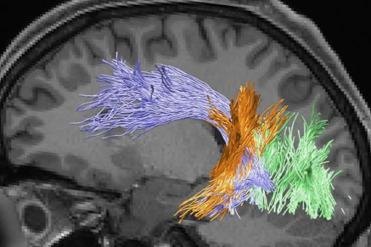
In this study, researchers in NICT and Osaka University examined the neuronal basis of individual differences in stereoacuity by combining cutting-edge neuroimaging techniques and psychophysics. We specifically focused on white matter pathways connecting distant brain areas because previous studies have demonstrated multiple brain areas in the dorsal and ventral visual areas are involved in stereopsis. We first identified trajectories of major white matter pathways related to visual processing by analyzing a diffusion MRI dataset. We then quantified neural tissues in the white matter using a quantitative MRI method. Furthermore, we estimated the stereoacuity of each individual human participant in a psychophysical experiment.
As a result, we found that the group with good stereoacuity showed significantly higher neural tissue density along a specific white matter pathway, the vertical occipital fasciculus (VOF) in the right hemisphere, compared to the group with poor stereoacuity. A functional MRI experiment revealed that the dorsal and ventral visual areas connected by the VOF are involved in stereopsis. Finally, we also confirmed that the neural tissue properties of the VOF did not correlate with performance in contrast detection, which does not require binocular integration of visual information. These results suggest that the communication between the dorsal and ventral visual brain areas via the VOF plays an important role in human stereopsis
A previous study found that approximately 30% of healthy individuals have relatively lower performance in stereoacuity. Further research on stereoacuity and white matter will contribute to a method for improving our performance in stereopsis.
Funding: The study was supported by the The Ministry of Internal Affairs and Communications.
Source: Sachiko Hirota – NICT
Publisher: Organized by NeuroscienceNews.com.
Image Source: NeuroscienceNews.com image is credited to NICT.
Original Research: Open access research for “Microstructural properties of the vertical occipital fasciculus explain the variability in human stereoacuity” by Hiroki Oishi, Hiromasa Takemura, Shuntaro C. Aoki, Ichiro Fujita, and Kaoru Amano in PNAS. Published October 1715 2018.
doi:10.1073/pnas.1804741115
[cbtabs][cbtab title=”MLA”]NICT”White Matter Pathway and Individual Variability in Human Stereoacuity.” NeuroscienceNews. NeuroscienceNews, 19 November 2018.
<https://neurosciencenews.com/stereoacuity-white-matter-10231/>.[/cbtab][cbtab title=”APA”]NICT(2018, November 19). White Matter Pathway and Individual Variability in Human Stereoacuity. NeuroscienceNews. Retrieved November 19, 2018 from https://neurosciencenews.com/stereoacuity-white-matter-10231/[/cbtab][cbtab title=”Chicago”]NICT”White Matter Pathway and Individual Variability in Human Stereoacuity.” https://neurosciencenews.com/stereoacuity-white-matter-10231/ (accessed November 19, 2018).[/cbtab][/cbtabs]
Abstract
Microstructural properties of the vertical occipital fasciculus explain the variability in human stereoacuity
Stereopsis is a fundamental visual function that has been studied extensively. However, it is not clear why depth discrimination (stereoacuity) varies more significantly among people than other modalities. Previous studies have reported the involvement of both dorsal and ventral visual areas in stereopsis, implying that not only neural computations in cortical areas but also the anatomical properties of white matter tracts connecting those areas can impact stereopsis. Here, we studied how human stereoacuity relates to white matter properties by combining psychophysics, diffusion MRI (dMRI), and quantitative MRI (qMRI). We performed a psychophysical experiment to measure stereoacuity and, in the same participants, we analyzed the microstructural properties of visual white matter tracts on the basis of two independent measurements, dMRI (fractional anisotropy, FA) and qMRI (macromolecular tissue volume; MTV). Microstructural properties along the right vertical occipital fasciculus (VOF), a major tract connecting dorsal and ventral visual areas, were highly correlated with measures of stereoacuity. This result was consistent for both FA and MTV, suggesting that the behavioral–structural relationship reflects differences in neural tissue density, rather than differences in the morphological configuration of fibers. fMRI confirmed that binocular disparity stimuli activated the dorsal and ventral visual regions near VOF endpoints. No other occipital tracts explained the variance in stereoacuity. In addition, the VOF properties were not associated with differences in performance on a different psychophysical task (contrast detection). These series of experiments suggest that stereoscopic depth discrimination performance is, at least in part, constrained by dorso-ventral communication through the VOF.