

Even when we’re resting, our brains are preparing us to be social, UCLA psychologists report.
A new study by UCLA neuroscientists sheds light on why Facebook is such a popular diversion for people who feel like taking a break. Their research shows that even during quiet moments, our brains are preparing us to be socially connected to other people.
“The brain has a major system that seems predisposed to get us ready to be social in our spare moments,” said Matthew Lieberman, a UCLA professor of psychology and of psychiatry and biobehavioral sciences. “The social nature of our brains is biologically based.”
The research helps resolve a nearly 20-year-old mystery. Neuroscientists have known since the 1990s that the brain includes a network of regions that seems to be most active during periods of rest — this became apparent when they examined brain scans of people who were attempting to answer challenging questions during scientific experiments and noticed that certain areas of the brain became unusually active during the periods in between the problem-solving. But until now, scientists knew very little about what purpose is served by the brain’s activity during those interludes.
The UCLA research, published in the June print edition of the Journal of Cognitive Neuroscience, shows that during quiet moments, the brain is preparing to focus on the minds of other people — or to “see the world through a social lens,” said Lieberman, the study’s senior author.
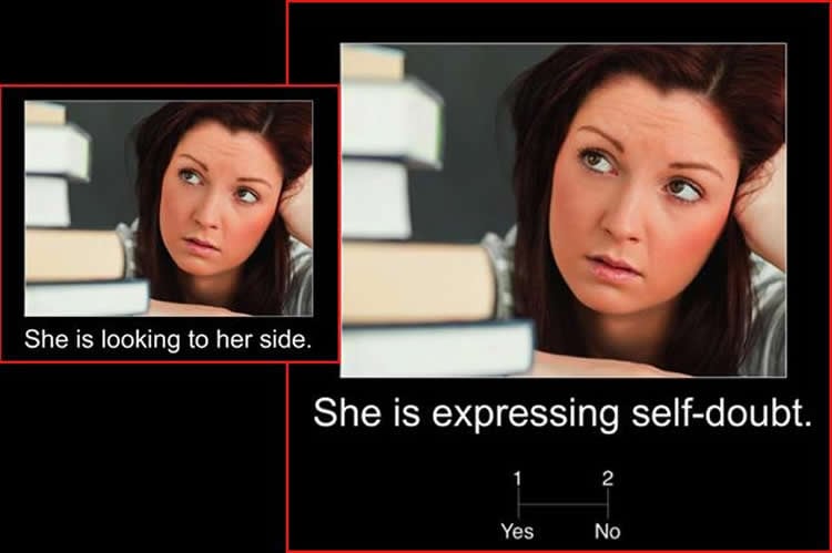
In experiments at UCLA’s Ahmanson-Lovelace Brain Mapping Center, the researchers showed photos with captions to 21 people, and tracked their brain activity using functional magnetic resonance imaging, or fMRI. Most of the photos showed people performing actions in a social setting and expressing a certain emotion. In one set of 40 photographs, images were paired with captions that reflected the person’s mental state — “He is feeling bored” or “She is expressing self-doubt,” for example. The second set of photos had identical images, but with captions that merely described what the person was doing — “He is resting his head” or “She is looking to her side.” And a third set of images depicted a number accompanied by a simple mathematical equation — for example, “10: 18-8.”
Participants were asked to judge whether the captions accurately expressed what the images showed. Among the findings:
- The same regions of the brain that were active during the brief times that subjects were not looking at photos were also active when the participants were considering the photos with captions about people’s emotions. But those areas of the brain were not active when the participants were viewing the cards with captions about the person’s physical activity and those with the math equations.
- Sometimes, a part of the brain called the dorsomedial prefrontal cortex was more active during the rest period immediately before participants were asked to look at photos. In those cases, the participants made significantly faster judgments if the next photo they saw presented a statement about the person’s mental state.
- There was no relation between activity in the dorsomedial prefrontal cortex during rest and the speed of people’s decision-making on the questions involving math equations or the photographs with physical descriptions.
- Study participants who were found to have traits characteristic of autism spectrum disorders — the researchers identified them using questionnaires administered prior to the brain scans — had less brain activity in the dorsomedial prefrontal cortex during periods of rest and were slower to judge the mental state of people in the photographs. Those with the least amount of dorsomedial prefrontal cortex activity were 10 percent slower than those with the most.
- The difference in decision-making speed that the researchers observed could have a significant effect in people’s everyday lives, Lieberman said. “It might not seem like a huge advantage, but being 10 percent faster, time after time, in each conversation will allow a person to be much better prepared and in control of their social lives.”
Lieberman, author of the best-selling book “Social: Why Our Brains Are Wired to Connect,” describes the dorsomedial prefrontal cortex as the “CEO of the social brain.” It’s part of a network in the brain that turns on when we dream and during periods of rest, in addition to when we explicitly think about other people, he said.
Based on activity in that region of the brain when the study participants were resting, researchers could accurately predict how quickly the participants would perform the next task. When the dorsomedial prefrontal cortex was highly active before participants saw a photo with a description of a mental state, they were faster in making their judgment; when the region was only slightly active, their decision-making was slower. The phenomenon applied equally among men and women.
“It’s the same photograph; the only thing that differs is whether the caption is mind-focused or body-focused,” said lead author Robert Spunt, who conducted the research when he was a UCLA doctoral student in psychology and is now a postdoctoral scholar at Caltech. “It’s remarkable.”
Lieberman said that people who struggle to read social cues in other people’s facial expressions might be able to improve this skill with practice, and he is conducting additional research to examine whether certain kinds of practice at social thinking can help improve people’s social abilities more broadly.
The findings suggest that the dorsomedial prefrontal cortex might turn on during dreams and rest in order to process our recent social experiences and update our assumptions and understanding of the social world, Lieberman said.
“It is getting us ready to see the world socially in terms of other people’s thoughts, feelings and goals,” he said. “That indicates it is important; the brain doesn’t just turn systems on. We walk around with our brain trying to reset itself to start thinking about other minds.”
So although Facebook might not have been designed with the dorsomedial prefrontal cortex in mind, the social network is very much in sync with how our brains are wired.
“When I want to take a break from work, the brain network that comes on is the same network we use when we’re looking through our Facebook timeline and seeing what our friends are up to,” said Lieberman, one of the founders of the field of study known as social cognitive neuroscience. “That’s what our brain wants to do, especially when we take a break from work that requires other brain networks.”
Source: Stuart Wolpert – UCLA
Image Source: The image is credited to Robert Spunt
Original Research: Full open access research for “The Default Mode of Human Brain Function Primes the Intentional Stance” by Robert P. Spunt, Meghan L. Meyer, and Matthew D. Lieberman in Journal of Cognitive Neuroscience. Published online April 30 2015 doi:10.1162/jocn_a_00785
Abstract
The Default Mode of Human Brain Function Primes the Intentional Stance
Humans readily adopt an intentional stance to other people, comprehending their behavior as guided by unobservable mental states such as belief, desire, and intention. We used fMRI in healthy adults to test the hypothesis that this stance is primed by the default mode of human brain function present when the mind is at rest. We report three findings that support this hypothesis. First, brain regions activated by actively adopting an intentional rather than nonintentional stance to a social stimulus were anatomically similar to those demonstrating default responses to fixation baseline in the same task. Second, moment-to-moment variation in default activity during fixation in the dorsomedial PFC was related to the ease with which participants applied an intentional—but not nonintentional—stance to a social stimulus presented moments later. Finally, individuals who showed stronger dorsomedial PFC activity at baseline in a separate task were generally more efficient when adopting the intentional stance and reported having greater social skills. These results identify a biological basis for the human tendency to adopt the intentional stance. More broadly, they suggest that the brain’s default mode of function may have evolved, in part, as a response to life in a social world.
“The Default Mode of Human Brain Function Primes the Intentional Stance” by Robert P. Spunt, Meghan L. Meyer, and Matthew D. Lieberman in Journal of Cognitive Neuroscience. Published online April 30 2015 doi:10.1162/jocn_a_00785


