

Summary: Slower walkers have accelerated aging in middle age, both physically and cognitively. Tests given to measure IQ, language, motor skills, and emotional control at age 3, can predict walking speed and thus accelerated aging during middle age.
Source: Duke Universtiy
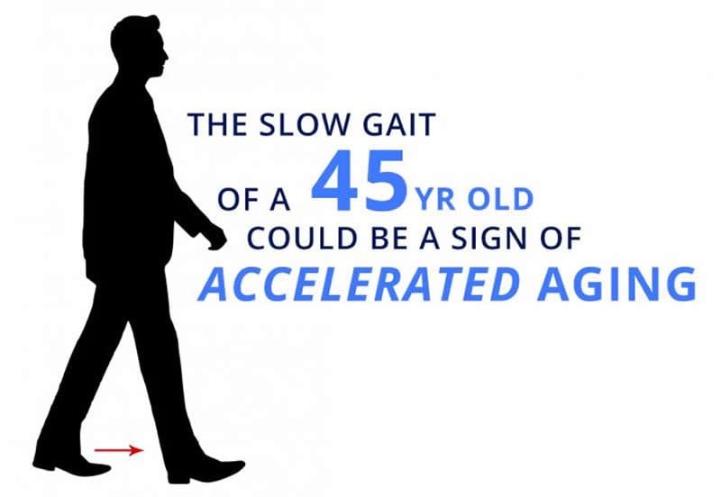
The walking speed of 45-year-olds, particularly their fastest walking speed without running, can be used as a marker of their aging brains and bodies.
Slower walkers were shown to have “accelerated aging” on a 19-measure scale devised by researchers, and their lungs, teeth and immune systems tended to be in worse shape than the people who walked faster.
“The thing that’s really striking is that this is in 45-year-old people, not the geriatric patients who are usually assessed with such measures,” said lead researcher Line J.H. Rasmussen, a post-doctoral researcher in the Duke University department of psychology & neuroscience.
Equally striking, neurocognitive testing that these individuals took as children could predict who would become the slower walkers. At age 3, their scores on IQ, understanding language, frustration tolerance, motor skills and emotional control predicted their walking speed at age 45.
“Doctors know that slow walkers in their seventies and eighties tend to die sooner than fast walkers their same age,” said senior author Terrie E. Moffitt, the Nannerl O. Keohane University Professor of Psychology at Duke University, and Professor of Social Development at King’s College London. “But this study covered the period from the preschool years to midlife, and found that a slow walk is a problem sign decades before old age.”
The data come from a long-term study of nearly 1,000 people who were born during a single year in Dunedin, New Zealand. The 904 research participants in the current study have been tested, quizzed and measured their entire lives, mostly recently from April 2017 to April 2019 at age 45.
The study appears Oct. 11 in JAMA Network Open.
MRI exams during their last assessment showed the slower walkers tended to have lower total brain volume, lower mean cortical thickness, less brain surface area and higher incidence of white matter “hyperintensities,” small lesions associated with small vessel disease of the brain. In short, their brains appeared somewhat older.
Adding insult to injury perhaps, the slower walkers also looked older to a panel of eight screeners who assessed each participant’s ‘facial age’ from a photograph.
Gait speed has long been used as a measure of health and aging in geriatric patients, but what’s new in this study is the relative youth of these study subjects and the ability to see how walking speed matches up with health measures the study has collected during their lives.
“It’s a shame we don’t have gait speed and brain imaging for them as children,” Rasmussen said. (The MRI was invented when they were five, but was not given to children for many years after.)
Some of the differences in health and cognition may be tied to lifestyle choices these individuals have made. But the study also suggests that there are already signs in early life of who would become the slowest walkers, Rasmussen said. “We may have a chance here to see who’s going to do better health-wise in later life.”
Funding: This research was supported by grants the US National Institute on Aging (AG032282, AG049789, AG028716), the UK Medical Research Council (MR/P005918/1), the Jacobs Foundation, the New Zealand Health Research Council (16-604), the New Zealand Ministry of Business, Innovation and Employment, the Lundbeck Foundation (R288-2018-380), the US National Science Foundation (NSF DGE-1644868), the US National Institute of Child Health and Human Development (T32-HD007376).
Source:
Duke Universtiy
Media Contacts:
Karl Leif Bates – Duke Universtiy
Image Source:
The image is credited to Duke University Communications.
Original Research: Open access
“”Association of Neurocognitive and Physical Function With Gait Speed in Midlife”. Line Rasmussen, Avshalom Caspi, Anthony Ambler, et al.
JAMA Network Open doi:10.1001/jamanetworkopen.2019.13123.
Abstract
“Association of Neurocognitive and Physical Function With Gait Speed in Midlife
Importance
Gait speed is a well-known indicator of risk of functional decline and mortality in older adults, but little is known about the factors associated with gait speed earlier in life.
Objectives
To test the hypothesis that slow gait speed reflects accelerated biological aging at midlife, as well as poor neurocognitive functioning in childhood and cognitive decline from childhood to midlife.
Design, Setting, and Participants
This cohort study uses data from the Dunedin Multidisciplinary Health and Development Study, a population-based study of a representative 1972 to 1973 birth cohort in New Zealand that observed participants to age 45 years (until April 2019). Data analysis was performed from April to June 2019.
Exposures
Childhood neurocognitive functions and accelerated aging, brain structure, and concurrent physical and cognitive functions in adulthood.
Main Outcomes and Measures
Gait speed at age 45 years, measured under 3 walking conditions: usual, dual task, and maximum gait speeds.
Results
Of the 1037 original participants (91% of eligible births; 535 [51.6%] male), 997 were alive at age 45 years, of whom 904 (90.7%) had gait speed measured (455 [50.3%] male; 93% white). The mean (SD) gait speeds were 1.30 (0.17) m/s for usual gait, 1.16 (0.23) m/s for dual task gait, and 1.99 (0.29) m/s for maximum gait. Adults with more physical limitations (standardized regression coefficient [β], −0.27; 95% CI, −0.34 to −0.21; P < .001), poorer physical functions (ie, weak grip strength [β, 0.36; 95% CI, 0.25 to 0.46], poor balance [β, 0.28; 95% CI, 0.21 to 0.34], poor visual-motor coordination [β, 0.24; 95% CI, 0.17 to 0.30], and poor performance on the chair-stand [β, 0.34; 95% CI, 0.27 to 0.40] or 2-minute step tests [β, 0.33; 95% CI, 0.27 to 0.39]; all P < .001), accelerated biological aging across multiple organ systems (β, −0.33; 95% CI, −0.40 to −0.27; P < .001), older facial appearance (β, −0.25; 95% CI, −0.31 to −0.18; P < .001), smaller brain volume (β, 0.15; 95% CI, 0.06 to 0.23; P < .001), more cortical thinning (β, 0.09; 95% CI, 0.02 to 0.16; P = .01), smaller cortical surface area (β, 0.13; 95% CI, 0.04 to 0.21; P = .003), and more white matter hyperintensities (β, −0.09; 95% CI, −0.15 to −0.02; P = .01) had slower gait speed. Participants with lower IQ in midlife (β, 0.38; 95% CI, 0.32 to 0.44; P < .001) and participants who exhibited cognitive decline from childhood to adulthood (β, 0.10; 95% CI, 0.04 to 0.17; P < .001) had slower gait at age 45 years. Those with poor neurocognitive functioning as early as age 3 years had slower gait in midlife (β, 0.26; 95% CI, 0.20 to 0.32; P < .001).
Conclusions and Relevance
Adults’ gait speed is associated with more than geriatric functional status; it is also associated with midlife aging and lifelong brain health.


