

Summary: A new oral device, dubbed the mandibular advancement appliance, can improve the quality of sleep for those with sleep apnea. The device increases retroglossal airway space three-dimensionally.
Source: Hiroshima University
Researchers measured a novel treatment for sleep apnea developed at Hiroshima University Hospital with positive results. By measuring patients lying down flat, the researchers stimulated sleep conditions and measured the patient’s airways using 3D imaging. The study confirmed that the treatment is effective at opening the airways and warrants further collaboration between dentists and doctors in the treatment of sleep apnea.
Obstructive sleep apnea is a condition that causes throat muscles to relax and narrows the airways of those affected while they are asleep. Snorting, choking or gasping while sleeping are the indicators of the condition. Usually, the sufferers’ partner notices before they do!
People with mild to moderate sleep apnea experience daily fatigue and a shortened attention span from lack of sleep. Sleep apnea can also have more serious consequences; people have died from very severe forms of the condition.
“Your eyes are closed but you’re not resting,” explains Dr. Cynthia Concepción-Medina, Research Assistant at the Department of Orthodontics at Hiroshima University Hospital who contributed to the study with her colleagues Associate Professor Hiroshi Ueda and Dr. Yu Matsumura.
Treatments include a continuous positive airway pressure (CPAP) machine (a mask worn by the patient that delivers air pressure throughout the night) or one-piece oral appliances.
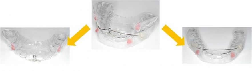
The Department of Orthodontics at Hiroshima University Hospital developed an oral appliance to help patients with mild to moderate sleep apnea. This appliance brings the jawbone forward to enlarge the air passageways at the back of their mouth. Each appliance is custom made for each patient and allows jaw movement, so it doesn’t affect the patient’s teeth or change the shape of their face.
“This is like when you have to use glasses, you have to wear them every time you want to see properly so [patients] have to wear this appliance every time [they] want to sleep better.” according to Dr. Ueda.
To further investigate how well the appliance works the research team, led by Dr. Matsumura, scanned a group of patients with mild-to-moderate sleep apnea using Multislice Computed Tomography (MSCT)– a type of X-ray where the machine rotates around an object, and it takes a picture each time it rotates. This data is then combined to see a 3D object and is a fast and precise method of scanning.
Previous research usually measured patients standing up, which does not simulate sleeping conditions. This study (published in Sleep Disorders, 2019) measured the change in airway space of 13 patients lying flat. The team found that the appliance had a positive effect on patients: wearing it almost halved the number of times the patients had sleep apnea episodes during the night and widened their airways to allow easier breathing.
“I think it’s unique research because we are dentists, but we can contribute to improving the [patient’s] sleep situation or breathing situation,” says Dr. Ueda.
This study indicates promising effects of this treatment and the team hopes that they can continue this collaboration between the dental and the medical field.
Source:
Hiroshima University
Media Contacts:
Norifumi Miyokawa – Hiroshima University
Image Source:
The image is credited to Department of Orthodontics and Craniofacial Developmental Biology/Hiroshima University Hospital.
Original Research: Open access.
“Multislice Computed Tomography Assessment of Airway Patency Changes Associated with Mandibular Advancement Appliance Therapy in Supine Patients with Obstructive Sleep Apnea”
Yu Matsumura, Hiroshi Ueda,Toshikazu Nagasaki, Cynthia Concepción Medina, Koji Iwai, and Kotaro Tanimoto Sleep Disorders
Volume 2019, Article ID 8509820, 9 pages doi:10.1155/2019/8509820
Abstract
Multislice Computed Tomography Assessment of Airway Patency Changes Associated with Mandibular Advancement Appliance Therapy in Supine Patients with Obstructive Sleep Apnea
The purpose of the present study was to measure the regional effects of the mandibular advancement appliance (MAA) on the upper airway of supine subjects with obstructive sleep apnea (OSA) using multislice computed tomography (MSCT). The subjects included 8 males and 5 females who were diagnosed with mild to moderate OSA and were referred to the Orthodontic Clinic of Hiroshima University Hospital, where they underwent MAA therapy. Using a CT scanner, baseline MSCT images were obtained from the subjects without the MAA for morphological analysis, and then the experimental images were obtained while wearing the MAA. To measure the anteroposterior diameter, width, and cross-sectional area of the oropharynx region of interest (ROI), five distance variables were first defined on each multiplanar reconstruction (MPR) image using OsiriX. Additionally, the volumes of the upper airway, bony hard tissue, and soft tissue (soft palate and tongue) in the oro-hypopharyngeal region were measured. In most of the assessed airway size variables, significant increases in the anteroposterior diameter and width were observed after MAA therapy. Regarding the upper airway cross-sectional area, all the upper airway size variables exhibited significant increases. In the volumetric analysis, a significant increase was observed in airway volume, whereas the soft tissue volume in the oro-hypopharyngeal region did not show the significant decrease after MMA therapy. However, from a different point of view, the volumes of the upper airway and soft tissue significantly increased and decreased, respectively, as demonstrated by the calculated ratio for the oro-hypopharyngeal region. We demonstrated that the proportional size of the soft tissue volume, i.e., the soft palate and tongue in the oro-hypopharyngeal region, significantly decreased during use of an MAA. This forward displacement of the soft tissue thereby increases the retroglossal airway space (except the nasopharynx) three-dimensionally.


