Summary: A new study implicated interneurons and pyramidal cells in the ability of a seizure to spread through the brain.
Source: University of Bonn
In some forms of epilepsy, the function of certain “brake cells” in the brain is presumed to be disrupted. This may be one of the reasons why the electrical malfunction is able to spread from the point of origin across large parts of the brain. A current study by the University of Bonn, in which researchers from Lisbon were also involved, points in this direction. The results will be published shortly in the renowned Journal of Neuroscience, but are already available online.
For their study, the researchers investigated rats suffering from temporal lobe epilepsy. This is the most common form of the disease in humans. Unfortunately, it barely responds to the currently available medicines. “This makes it all the more important to determine exactly how it arises,” stresses Dr. Leonie Pothmann, who completed her doctorate on the subject at the Institute of Experimental Epileptology at the University of Bonn.
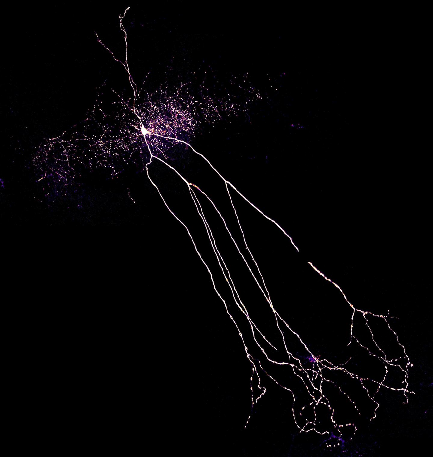
The data that has just been published may help scientists with this endeavor, because they indicate that a certain cell type does not function properly in patients. The affected cells are a class of so-called inhibitory interneurons, which are cells that can attenuate the excitation of brain areas. “We investigated interneurons in the hippocampus, an area of the temporal lobe known as the focus of epileptic seizures,” explains Pothmann.
Pyramidal cells play an important role in the transmission of excitation in the hippocampus. They generate voltage pulses in response to an electrical stimulus. These stimulate, among other things, interneurons, which in turn inhibit the pyramidal cells. This feedback loop acts as a kind of brake: It prevents the voltage pulses from propagating unhindered. An epileptic seizure would thus be nipped in the bud before it is able to spread to other parts of the brain.
Brake simulation in the computer
“In the rats, however, this brake did not work well compared to healthy animals,” says Pothmann’s colleague Dr. Oliver Braganza. “Our measurements show that the rapid, robust inhibition that occurs in healthy animals is greatly reduced in sick animals.”
In order to find out why this might happen and what the effects might be, the scientists simulated the interaction of pyramidal cell and interneuron on the computer. They changed certain properties of the virtual interneuron until its behavior in the simulation was exactly the same as in the sick animals.
The results provide information about two possible disturbances: The interneurons appear to release only a small part of the signal molecules (neurotransmitters) stored inside their cells in response to a stimulus. Additionally, their membranes are not working properly: They are unable to maintain a voltage gradient very well, almost as if they had a slight short circuit. Both factors contribute to the interneurons being activated only relatively weakly. In computer simulation, this interaction with the pyramidal cells resulted in the unhindered transmission of the type of activity that occurs in epileptic seizures.
“We now have to investigate these findings further,” explains Prof. Dr. Heinz Beck, head of the Institute of Experimental Epileptology and Associate Member of the German Center for Neurodegenerative Diseases. “First we have to find out whether the two disruptions are actually responsible for the malfunction of the interneurons. If so, this may open the way to new therapeutic approaches in the long term.” However, the results are still pure basic research, he emphasizes. “It is by no means clear whether they will benefit patients – and if they do, it will certainly take many more years.”
Source:
University of Bonn
Media Contacts:
Dr. Heinz Beck – University of Bonn
Image Source:
The image is credited to Leonie Pothmann/Uni Bonn.
Original Research: Open access
“Altered dynamics of canonical feed-back inhibition predicts increased burst transmission in chronic epilepsy”. Leonie Pothmann, Christian Klos, Oliver Braganza, Sarah Schmidt, Oihane Horno, Raoul-Martin Memmesheimer and Heinz Beck.
Journal of Neuroscience doi:10.1523/JNEUROSCI.2594-18.2019.
Abstract
Altered dynamics of canonical feed-back inhibition predicts increased burst transmission in chronic epilepsy
Inhibitory interneurons, organized into canonical feed-forward and feed-back motifs, play a key role in controlling normal and pathological neuronal activity. We demonstrate prominent quantitative changes in the dynamics of feed-back inhibition in a rat model of chronic epilepsy (male Wistar rats). Systematic interneuron recordings revealed a large decrease in intrinsic excitability of basket cells and OLM interneurons in epileptic animals. Additionally, the temporal dynamics of interneuron recruitment by recurrent feed-back excitation was strongly altered, resulting in a profound loss of initial feed-back inhibition during synchronous CA1 pyramidal activity. Biophysically constrained models of the complete feed-back circuit motifs of normal and epileptic animals revealed that as a consequence of altered feed-back inhibition, burst activity arising in CA3 is more strongly converted to a CA1 output. This suggests that altered dynamics of feed-back inhibition promote the transmission of epileptiform bursts to hippocampal projection areas.
SIGNIFICANCE STATEMENT
We quantitatively characterized changes of the CA1 feedback inhibitory circuit in a model of chronic temporal lobe epilepsy. This study shows for the first time that dynamic recruitment of inhibition in feedback circuits is altered and establishes the cellular mechanisms for this change. Computational modelling revealed that the observed changes are likely to systematically alter CA1 input-output properties leading to i) increased seizure propagation through CA1 and ii) altered computation of synchronous CA3 input.