

Summary: A new study reveals how patterns of neural activity in the retrosplenial cortex assist with navigation and spatial memory.
Source: VIB Flanders.
Navigation in mammals including humans and rodents depends on specialized neural networks that encode the animal’s location and trajectory in the environment, serving essentially as a GPS, findings that led to the 2014 Nobel Prize in Medicine. Failure of these networks to function properly, as seen in Alzheimer’s disease and other neurological conditions, results in severe disorientation and memory deficits. Researchers at NERF (VIB-imec-KU Leuven) have now uncovered striking neural activity patterns in a brain area called the retrosplenial cortex that may assist with spatial memory and navigation.
The prime example of spatial information coding is the firing of so called place cells in the hippocampus, a brain area known for its role in navigation and memory formation. Place cells fire when an animal enters a specific place in its environment. At any given location, only a small fraction of place cells is active, leaving the remaining neurons largely silent. This sparse firing pattern maximizes information storage in memory networks, but at the same time minimizes energy demands.
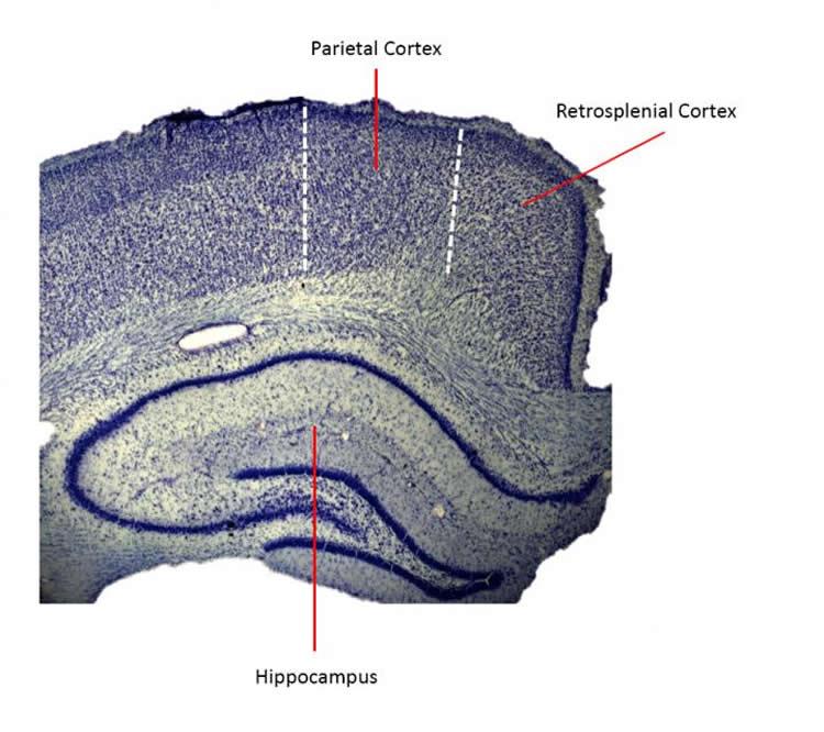
The hippocampus, however, is not the only brain area involved in spatial orientation and learning. The retrosplenial cortex is also highly active during navigation and memory retrieval and connects the hippocampus to the visual cortex and other areas of the brain. Damage to the retrosplenial cortex results in memory deficits and disorientation, and patients with Alzheimer’s disease have reduced activity in their retrosplenial cortex.
To better understand the role of the retrosplenial cortex, Drs. Dun Mao and Steffen Kandler, researchers in the laboratories of Profs. Vincent Bonin and Bruce McNaughton at NERF, measured its activity in mice that moved on a treadmill fitted with tactile stimuli. In this setting they could precisely track the animal’s behavior and location. By combining genetic labeling of cortical neurons and highly sensitive live microscopic techniques, the researchers were able to compare the activity of the neurons in the retrosplenial cortex with those in the hippocampus.
“Previous studies could only record from a few retrosplenial neurons simultaneously. With our cellular imaging technique, we could monitor the activity of hundreds to thousands of neurons simultaneously, which gave us a rich view into the neurons’ activity patterns,” explains prof. Vincent Bonin.
The researchers discovered a new group of cells that fire in smooth sequences as the animals run in the environment. Their activity resembled that of hippocampal place cells in terms of their sparse firing properties; however, the retrosplenial neurons responded differently to sensory inputs.
These results indicate that the retrosplenial cortex carries rich spatial activity, the mechanisms of which may be partially different from that of the hippocampus. They pave the way for a better understanding of how our brain processes spatial information. Prof. Vincent Bonin: “The next step is to investigate directly the relationship between retrosplenial activity and hippocampus as well as its link to visual inputs. It will also be interesting to know how activity in the retrosplenial cortex relates to the development of different neuronal diseases in mouse models.”
The study is an international collaboration between the laboratories of Dr. Vincent Bonin at NERF and Dr. Bruce McNaughton at the University of Lethbridge, Canada. Dr. Bonin is Principal investigator at NERF and VIB since 2011 and Associate Professor at KU Leuven since 2017 (Assistant since 2012). Dr. McNaughton is AHFMR/AIHS Polaris Research Chair at the Canadian Centre for Behavioural Neuroscience at The University of Lethbridge, and Distinguished Professor of Neurobiology and Behavior at UC Irvine. Dr. McNaughton held a Senior Visiting Scientist position at NERF from 2010 to 2013. The work was supported by core grants from imec, VIB and KU Leuven and from the Government of Flanders as well as research awards from FWO, Alberta Innovates: Health Solutions, NSERC, and NSF.
Source: Sooike Stoops – VIB Flanders
Image Source: NeuroscienceNews.com image is credited to Andrew Alexander and Douglas Nitz, UC San Diego.
Original Research: Full open access research for “Sparse orthogonal population representation of spatial context in the retrosplenial cortex” by Dun Mao, Steffen Kandler, Bruce L. McNaughton & Vincent Bonin in Nature Communications. Published online August 15 2017 doi:10.1038/s41467-017-00180-9
[cbtabs][cbtab title=”MLA”]VIB Flanders “New Brain Area Implicated in Navigation and Spatial Memory.” NeuroscienceNews. NeuroscienceNews, 16 August 2017.
<https://neurosciencenews.com/retrosplenial-cortex-navigation-7316/>.[/cbtab][cbtab title=”APA”]VIB Flanders (2017, August 16). New Brain Area Implicated in Navigation and Spatial Memory. NeuroscienceNew. Retrieved August 16, 2017 from https://neurosciencenews.com/retrosplenial-cortex-navigation-7316/[/cbtab][cbtab title=”Chicago”]VIB Flanders “New Brain Area Implicated in Navigation and Spatial Memory.” https://neurosciencenews.com/retrosplenial-cortex-navigation-7316/ (accessed August 16, 2017).[/cbtab][/cbtabs]
Abstract
Sparse orthogonal population representation of spatial context in the retrosplenial cortex
Sparse orthogonal coding is a key feature of hippocampal neural activity, which is believed to increase episodic memory capacity and to assist in navigation. Some retrosplenial cortex (RSC) neurons convey distributed spatial and navigational signals, but place-field representations such as observed in the hippocampus have not been reported. Combining cellular Ca2+ imaging in RSC of mice with a head-fixed locomotion assay, we identified a population of RSC neurons, located predominantly in superficial layers, whose ensemble activity closely resembles that of hippocampal CA1 place cells during the same task. Like CA1 place cells, these RSC neurons fire in sequences during movement, and show narrowly tuned firing fields that form a sparse, orthogonal code correlated with location. RSC ‘place’ cell activity is robust to environmental manipulations, showing partial remapping similar to that observed in CA1. This population code for spatial context may assist the RSC in its role in memory and/or navigation.
“Sparse orthogonal population representation of spatial context in the retrosplenial cortex” by Dun Mao, Steffen Kandler, Bruce L. McNaughton & Vincent Bonin in Nature Communications. Published online August 15 2017 doi:10.1038/s41467-017-00180-9


