

Summary: Researchers report that following amputation of one limb, the bioelectrical properties are immediately reflected in the undamaged, opposing limb.
Source: Tufts University.
In research that extends knowledge about the physiology of regeneration and wound repair, Tufts University biologists have discovered that amputation of one limb is immediately reflected in the bioelectric properties of the contralateral, or opposing, un-damaged limb of developing frogs. The pattern of bioelectric depolarization in the un-injured leg is directly correlated to the position and type of injury, indicating that information about damage to tissues is available to their symmetrical counterparts within about 30 seconds of injury. The newly discovered phenomenon, dubbed “bioelectric injury mirroring” or BIM, is described in detail in a paper to be published in the journal Development.
Bioelectric phenomena are caused by cells that produce a voltage potential across their membrane by actively pumping or passively diffusing charged ions into or out of the cell. Most cells are capable of doing this, and patterns of high and low voltage potentials help direct the proliferation and differentiation of cells, as well as the patterning of tissues and organs, during embryonic development. Bioelectric states have also been implicated in regeneration — researchers have been able to alter bioelectric state to induce the regeneration of tails on tadpoles that had already matured beyond the capability of regeneration.
Studies of the bioelectric contribution to regeneration have largely focused on the region around the wound. “However, we wanted to look a little further,” said Sera Busse, the study’s first author who conducted the research as an undergraduate student before graduating with a degree in biology from Tufts in May 2018. “We know that bioelectric potentials can exhibit symmetrical patterns. We asked whether it was possible that the patterns resulting from injury might also be reflected symmetrically, at a distance.”
Busse, post-doctoral scholar Patrick McMillen, Ph.D., and Michael Levin, Ph.D., the Vannevar Bush Professor of Biology in the School of Arts & Sciences and Director of the Allen Discovery Center at Tufts, devised experiments using a fluorescent dye that can reveal the pattern of electrical depolarization in the upper layer of skin.
When Busse amputated the limbs of froglets still in regeneration stage, the dye revealed a remarkable phenomenon: The un-injured leg exhibited bioelectric states that mirrored the location and type of injury occurring on the opposite side, and the effect was immediate, occurring within 5 seconds.
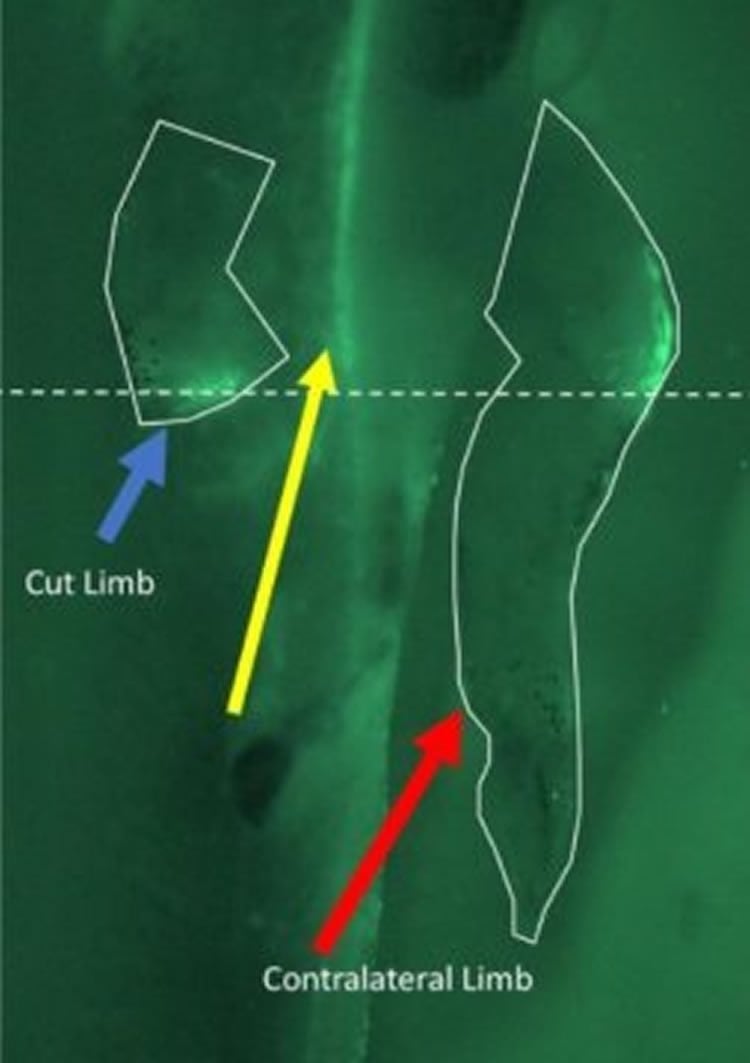
“What was amazing about this result was that not only did the depolarization in the un-injured leg detect the presence of injury on the other side, it also reflected information about the position of the cut,” said Levin.
The researchers considered whether such information was conveyed by typical neural communication through the central nervous system or spinal cord, but the BIM signal was undiminished when the central nervous system communication was interrupted. The result suggests that the distant communication between limbs occurs by a cell-to-cell mechanism which may be an evolutionary precursor to the more familiar neural signaling.
“Looking ahead, we will be employing more precise genetically-encoded voltage sensing tools, which can provide more quantitative and deeper tissue information than dyes, and machine learning methods to extract signatures of different types of damage from the bioelectric signal, to provide a more highly resolved understanding of the BIM phenomenon,” said Levin. Next steps involve understanding the mechanism and information content of such long-range signaling in the body, and potentially developing surrogate-site diagnostics for many different disease conditions.
Funding: This research was supported by the Allen Discovery Center program through The Paul G. Allen Frontiers Group (grant #12171), and the W. M. Keck Foundation (grant #5903).
In addition to conducting leading research during her time as a Tufts undergraduate, Busse is a nationally competitive rower, a 10-time U.S. rock climbing team member and a U.S. national champion in the sport. The other two authors, McMillen and Levin, also are former undergraduates of Tufts University.
Source: Tufts University
Publisher: Organized by NeuroscienceNews.com.
Image Source: NeuroscienceNews.com image is credited to Sera Busse, Patrick McMillen, Michael Levin — Tufts University.
Original Research: Abstract for “Cross-limb communication during Xenopus hind-limb regenerative response: non-local bioelectric injury signals” by Sera M. Busse, Patrick T. McMillen, and Michael Levin in Development. Published October 5 2018.
doi:10.1242/dev.164210
[cbtabs][cbtab title=”MLA”]Tufts University”Amputation Injury is Communicated to Opposing Limbs.” NeuroscienceNews. NeuroscienceNews, 5 October 2018.
<https://neurosciencenews.com/remapping-amputation-9966/>.[/cbtab][cbtab title=”APA”]Tufts University(2018, October 5). Amputation Injury is Communicated to Opposing Limbs. NeuroscienceNews. Retrieved October 5, 2018 from https://neurosciencenews.com/remapping-amputation-9966/[/cbtab][cbtab title=”Chicago”]Tufts University”Amputation Injury is Communicated to Opposing Limbs.” https://neurosciencenews.com/remapping-amputation-9966/ (accessed October 5, 2018).[/cbtab][/cbtabs]
Abstract
Cross-limb communication during Xenopus hind-limb regenerative response: non-local bioelectric injury signals
Regeneration of damaged body-parts requires coordination of size, shape, location, and orientation of tissue with the rest of the body. It is not currently known how far injury sites communicate with the remaining soma during repair, or what information may emanate from the injury site to other regions. We examined the bioelectric properties (resting potential gradients in the epidermis) of Xenopus froglets undergoing hind-limb amputation and observed that the contralateral (un-damaged) limb exhibits apparent depolarization signals immediately after the opposite hind-limb is amputated. The pattern of depolarization matches that of the amputated limb and is correlated to the position and type of injury, revealing that information about damage is available to remote body tissues and is detectable non-invasively in vivo by monitoring of the bioelectric state. These data extend knowledge about the electrophysiology of regenerative response, identify a novel communication process via long-range spread of injury signaling, a phenomenon which we call bioelectric injury mirroring (BIM), and suggests revisions to regenerative medicine and diagnostic strategies focused entirely on the wound site and to the use of contralateral limbs as controls.


