Summary: Researchers report they have successfully linked a rabbit retina to a chip in vitro.
Source: Swiss National Science Foundation.
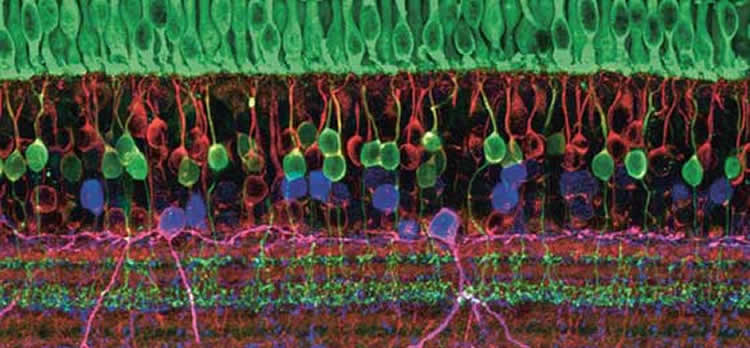
Researchers have linked a rabbit retina to a chip in vitro. It connected to thousands of transistors and helps us to understand how neurons process information.
Nystagmus is a genetically transmitted disease that causes an uncontrolled, back-and-forth twitching of the eyeball. Roughly one in every 1,500 men suffer from it. But before now, we did not know that this twitching is caused by retinal neurons making miscalculations when converting visual stimuli into electrical signals.
How does the brain – to which the retina also belongs – understand what information is contained in a stimulus? Until now, it was known that neurons answer stimuli by firing off salvos of electrical signals that are transmitted via synapses to other nerve cells. The information about the stimulus is actually contained in the number of impulses and in the time intervals between them. But just how these codes are read and written is still a subject of debate among neuroscientists.
Useful noise
Felix Franke from the Bioengineering Laboratory of ETH Zurich has come one step closer to understanding this process – at least as far as the retina is concerned. In a recently published study in the specialist journal Neuron, Franke’s team investigates whether it is more useful for the brain to ‘hear’ a whole orchestra of neurons at the same time, or only individual nerve cells. They found that if the brain hears them all, then it can learn more about the stimulus that caused the impulses – such as a picture that the eye has just seen.
In their experiment, Franke’s team linked the retinas of rabbits, laid out flat, with a computer chip containing 11,000 densely packed electrodes. They then moved a bright bar past the retinas. The neuroscientists were able to record the signals in the photoreceptors as data, via the electrodes.
The problem is that the nerve cells often answer differently to the same stimulus, which makes it difficult to form conclusions about the original stimulus. Franke explains this so-called ‘signal noise’ by using a dice metaphor: “If the stimulus is the number three, then one neurone will perhaps give us a two, and the neurone next to it a four. If we take the average of them both, the answer is correct. Viewed individually, each answer would be incorrect”. The ‘orchestra’ is thus more precise than individual neurons.
This has been confirmed by the neuroscientist Felipe Gerhard, who completed his doctorate at EPFL and is currently involved in research at Brown University in Providence, USA. The experiments with the rabbit retinas help him to process patterns in this signal noise, enabling him to recognise visual stimuli better. These findings should be a solid basis for future research on the neural code.
But this random signal noise in the brain can sometimes also hinder communication between neurons, says Gerhard: “Evolution has found ways of dealing with this noise, and even of using it”. It’s especially useful for creative thinking, he maintains.
Prostheses with a tactile sense
Franke believes that these findings might in future be employed for therapeutic purposes. “If we can understand how neural networks function, then we can also better understand the diseases that are connected with them”. Such as the nystagmus mentioned above.
Franke was involved in a study published in early 2016 that for the first-ever time found a connection between nystagmus in a human eye and a malformation of the retina in mice. It was the first time, says Franke, that a neural calculation was recognised as a factor in a human disease.
At Brown University, Gerhard also sees the possibility of therapeutic applications – such as prostheses controlled by thoughts. Arm prostheses could possibly even ‘write’ in the neural network of the brain and thereby recreate a sense of touch.
Currently, Gerhard is working with epilepsy patients measuring and analysing their neural activity during epileptic fits. Here, too, patterns in the signal noise play a role: “This could allow us to predict epileptic fits. As soon as the fit begins, we could attempt to stimulate these neurons actively, and thereby suppress the abnormal pattern”.
Source: Martin Angler – Swiss National Science Foundation
Image Source: This NeuroscienceNews.com image is adapted from the Swiss National Science Foundation press release.
Original Research: Abstract for “Structures of Neural Correlation and How They Favor Coding” by Felix Franke, Michele Fiscella, Maksim Sevelev, Botond Roska, Andreas Hierlemann, and Rava Azeredo da Silveira in Neuron. Published online January 20 2016 doi:10.1016/j.neuron.2015.12.037
[cbtabs][cbtab title=”MLA”]Swiss National Science Foundation. “New Clue to How Lithium Works in the Brain.” NeuroscienceNews. NeuroscienceNews, 8 July 2016.
<https://neurosciencenews.com/rabbit-retina-in-vitro-chip-4645/>.[/cbtab][cbtab title=”APA”]Swiss National Science Foundation. (2016, July 8). New Clue to How Lithium Works in the Brain. NeuroscienceNews. Retrieved July 8, 2016 from https://neurosciencenews.com/rabbit-retina-in-vitro-chip-4645/[/cbtab][cbtab title=”Chicago”]Swiss National Science Foundation. “New Clue to How Lithium Works in the Brain.” https://neurosciencenews.com/rabbit-retina-in-vitro-chip-4645/ (accessed July 8, 2016).[/cbtab][/cbtabs]
Abstract
Structures of Neural Correlation and How They Favor Coding
Highlights
•Neural noise correlations have stimulus-dependent structure in retina and cortex
•Analysis of neural data demonstrates that this structure improves visual coding
•Response models capture the emergence of this structure from circuit properties
•Neural coding models indicate that it can affect coding strategies qualitatively
Summary
The neural representation of information suffers from “noise”—the trial-to-trial variability in the response of neurons. The impact of correlated noise upon population coding has been debated, but a direct connection between theory and experiment remains tenuous. Here, we substantiate this connection and propose a refined theoretical picture. Using simultaneous recordings from a population of direction-selective retinal ganglion cells, we demonstrate that coding benefits from noise correlations. The effect is appreciable already in small populations, yet it is a collective phenomenon. Furthermore, the stimulus-dependent structure of correlation is key. We develop simple functional models that capture the stimulus-dependent statistics. We then use them to quantify the performance of population coding, which depends upon interplays of feature sensitivities and noise correlations in the population. Because favorable structures of correlation emerge robustly in circuits with noisy, nonlinear elements, they will arise and benefit coding beyond the confines of retina.
“Structures of Neural Correlation and How They Favor Coding” by Felix Franke, Michele Fiscella, Maksim Sevelev, Botond Roska, Andreas Hierlemann, and Rava Azeredo da Silveira in Neuron. Published online January 20 2016 doi:10.1016/j.neuron.2015.12.037