

Summary: Pyramidal cells in the CA2 region of the hippocampus are responsible for storing critical timing information.
Source: MIT
When we experience a new event, our brain records a memory of not only what happened, but also the context, including the time and location of the event. A new study from MIT neuroscientists sheds light on how the timing of a memory is encoded in the hippocampus, and suggests that time and space are encoded separately.
In a study of mice, the researchers identified a hippocampal circuit that the animals used to store information about the timing of when they should turn left or right in a maze. When this circuit was blocked, the mice were unable to remember which way they were supposed to turn next. However, disrupting the circuit did not appear to impair their memory of where they were in space.
The findings add to a growing body of evidence suggesting that when we form new memories, different populations of neurons in the brain encode time and place information, the researchers say.
“There is an emerging view that ‘place cells’ and ‘time cells’ organize memories by mapping information onto the hippocampus. This spatial and temporal context serves as a scaffold that allows us to build our own personal timeline of memories,” says Chris MacDonald, a research scientist at MIT’s Picower Institute for Learning and Memory and the lead author of the study.
Susumu Tonegawa, the Picower Professor of Biology and Neuroscience at the RIKEN-MIT Laboratory of Neural Circuit Genetics at the Picower Institute, is the senior author of the study, which appears this week in the Proceedings of the National Academy of Sciences.
Time and place
About 50 years ago, neuroscientists discovered that the brain’s hippocampus contains neurons that encode memories of specific locations. These cells, known as place cells, store information that becomes part of the context of a particular memory.
The other critical piece of context for any given memory is the timing. In 2011, MacDonald and the late Howard Eichenbaum, a professor of psychological and brain sciences at Boston University, discovered cells that keep track of time, in a part of the hippocampus called CA1.
In that study, MacDonald, who was then a postdoc at Boston University, found that these cells showed specific timing-related firing patterns when mice were trained to associate two stimuli — an object and an odor — that were presented with a 10-second delay between them. When the delay was extended to 20 seconds, the cells reorganized their firing patterns to last 20 seconds instead of 10.
“It’s almost like they’re forming a new representation of a temporal context, much like a spatial context,” MacDonald says. “The emerging view seems to be that both place and time cells organize memory by mapping experience to a representation of context that is defined by time and space.”
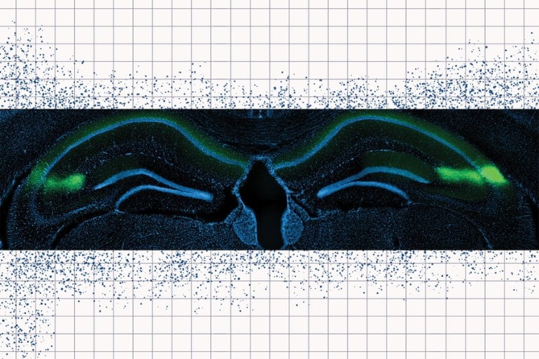
In the new study, the researchers wanted to investigate which other parts of the brain might be feeding CA1 timing information. Some previous studies had suggested that a nearby part of the hippocampus called CA2 might be involved in keeping track of time. CA2 is a very small region of the hippocampus that has not been extensively studied, but it has been shown to have strong connections to CA1.
To study the links between CA2 and CA1, the researchers used an engineered mouse model in which they could use light to control the activity of neurons in the CA2 region. They trained the mice to run a figure-eight maze in which they would earn a reward if they alternated turning left and right each time they ran the maze. Between each trial, they ran on a treadmill for 10 seconds, and during this time, they had to remember which direction they had turned on the previous trial, so they could do the opposite on the upcoming trial.
When the researchers turned off CA2 activity while the mice were on the treadmill, they found that the mice performed very poorly at the task, suggesting that they could no longer remember which direction they had turned in the previous trial.
“When the animals are performing normally, there is a sequence of cells in CA1 that ticks off during this temporal coding phase,” MacDonald says. “When you inhibit the CA2, what you see is the temporal coding in CA1 becomes less precise and more smeared out in time. It becomes destabilized, and that seems to correlate with them also performing poorly on that task.”
Memory circuits
When the researchers used light to inhibit CA2 neurons while the mice were running the maze, they found little effect on the CA1 “place cells” that allow the mice to remember where they are. The findings suggest that spatial and timing information are encoded preferentially by different parts of the hippocampus, MacDonald says.
“One thing that’s exciting about this work is this idea that spatial and temporal information can operate in parallel and might merge or separate at different points in the circuit, depending on what you need to accomplish from a memory standpoint,” he says.
MacDonald is now planning additional studies of time perception, including how we perceive time under different circumstances, and how our perception of time influences our behavior. Another question he hopes to pursue is whether the brain has different mechanisms for keeping track of events that are separated by seconds and events that are separated by much longer periods of time.
“Somehow the information that we store in memory preserves the sequential order of events across very different timescales, and I’m very interested in how it is that we’re able to do that,” he says.
Funding: The research was funded by the RIKEN Center for Brain Science, the Howard Hughes Medical Institute, and the JPB Foundation.
About this neuroscience research news
Source: MIT
Contact: Anne Trafton – MIT
Image: The image is credited to Tonegawa lab
Original Research: Closed access.
“Crucial role for CA2 inputs in the sequential organization of CA1 time cells supporting memory” by Christopher J. MacDonald and Susumu Tonegawa. PNAS
Abstract
Crucial role for CA2 inputs in the sequential organization of CA1 time cells supporting memory
There is considerable evidence for hippocampal time cells that briefly activate in succession to represent the temporal structure of memories. Previous studies have shown that time cells can be disrupted while leaving place cells intact, indicating that spatial and temporal information can be coded in parallel. However, the circuits in which spatial and temporal information are coded have not been clearly identified. Here we investigated temporal and spatial coding by dorsal hippocampal CA1 (dCA1) neurons in mice trained on a classic spatial working-memory task. On each trial, the mice approached the same choice point on a maze but were trained to alternate between traversing one of two distinct spatial routes (spatial coding phase). In between trials, there was a 10-s mnemonic delay during which the mouse continuously ran in a fixed location (temporal coding phase). Using cell-type–specific optogenetic methods, we found that inhibiting dorsal CA2 (dCA2) inputs into dCA1 degraded time cell coding during the mnemonic delay and impaired the mouse’s subsequent memory-guided choice. Conversely, inhibiting dCA2 inputs during the spatial coding phase had a negligible effect on place cell activity in dCA1 and no effect on behavior. Collectively, our work demonstrates that spatial and temporal coding in dCA1 is largely segregated with respect to the dCA2–dCA1 circuit and suggests that CA2 plays a critical role in representing the flow of time in memory within the hippocampal network.


