

Summary: A recent study in PNAS implicates alterations in the brain’s glutamate signaling in people with PTSD. Neuroimaging revealed increased levels of mGluR5 in those with post traumatic stress disorder. Researchers report drugs that can reduce mGluR5 function could help reduce fear, stress and other symptoms of PTSD.
Source: Yale.
A study of post-traumatic stress disorder (PTSD) — conducted by the VA National Center for PTSD (NCPTSD), National PTSD Brain Bank, and Yale University — has identified a new potential mechanism contributing to the biology of the disorder that may be targeted by future treatments.
Among combat veterans, PTSD is a common and disabling condition that is associated with high suicide risk, and in some cases it is difficult to treat effectively. Patients — civilians with significant trauma history and veterans with combat-related or civilian trauma history — are commonly treated with a combination of psychological therapy and medications aimed at alleviating diverse symptoms, such as hyper-arousal and depression.
At present, there are two medications approved by the FDA for the treatment of PTSD symptoms, but the limited effectiveness of these medications results in patients being treated frequently with multiple medications that are not specifically approved for PTSD, note the researchers.
“We really need to examine what is happening at a molecular level so we can start developing novel efficacious therapies,” said Sophie Holmes, postdoctoral research associate in the Yale Department of Psychiatry and lead author of the study published in the Proceedings of the National Academy of Sciences.
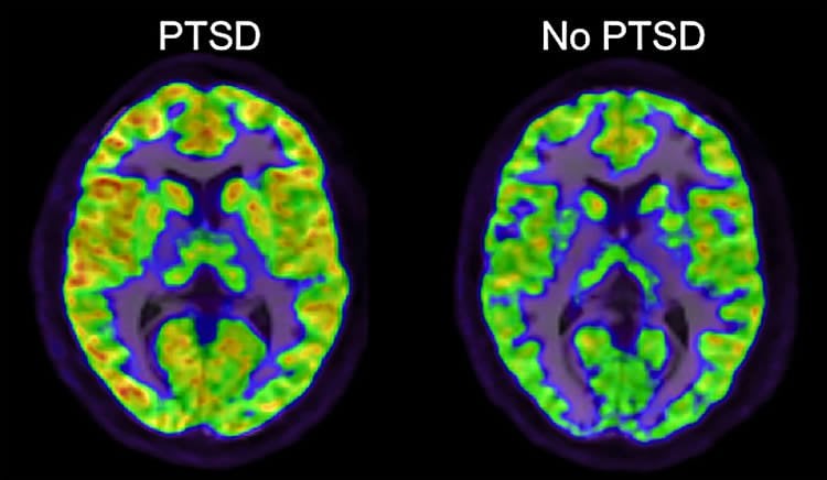
The study, led by NCPTSD and Yale psychiatrist Irina Esterlis, is the first to implicate a specific alteration in brain glutamate signaling in PTSD. Glutamate is a chemical messenger of brain signals, and alterations in glutamate levels in PTSD were described previously. The new study reports that positron emission tomography (PET) scans show increased levels of a subtype of glutamate receptor in the brain, metabotropic glutamate receptor-5 (mGluR5), in patients with PTSD. In animals, overstimulation of mGluR5 is associated with fear and stress-related behaviors; drugs that reduce mGluR5 function may reduce these symptoms. Thus, the current study may have implications for the treatment of PTSD, said the researchers.
This study also provided potential insights into how the increases in mGluR5 might arise. A novel and important feature of this study, according to the researchers, is that it is the first to link brain chemistry findings in patients with PTSD, as measured through PET scans, to detailed molecular analyses of brain changes in PTSD that can only be conducted in brain tissue that has been donated by veterans or their families for research purposes. The analyses, conducted in the laboratory of Yale psychiatrist Ronald Duman, found a clue at the level of gene expression, or the conversion of DNA to RNA. Messenger RNA (mRNA) levels of mGluR5 were not increased, but mRNA levels that code for the Shank1 protein were increased. Shank1 “activates” mGluR5 by linking them to the cell surface. The PET scans may have detected higher mGluR5 availability at the cell surface in PTSD.
“This study is one of the first examples to highlight the importance of the new National PTSD Brain Bank in enabling us to study the biology of PTSD at a deeper level. We have a long way to go to understand the complex neurobiology of PTSD, but these new research approaches should have a significant impact on the field”, said Esterlis.
Individuals or families seeking information about the National PTSD Brain Bank may visit its website or call 800-762-6609.
Source: Bill Hathaway – Yale
Image Source: NeuroscienceNews.com image is adapted from the Yale news release.
Original Research: Abstract for “Altered metabotropic glutamate receptor 5 markers in PTSD: In vivo and postmortem evidence” by Sophie E. Holmes, Matthew J. Girgenti, Margaret T. Davis, Robert H. Pietrzak, Nicole DellaGioia, Nabeel Nabulsi, David Matuskey, Steven Southwick, Ronald S. Duman, Richard E. Carson, John H. Krystal, Irina Esterlis, and the Traumatic Stress Brain Study Group in PNAS. Published online July 17 2017 doi:10.1073/pnas.1701749114
[cbtabs][cbtab title=”MLA”]Yale “Potential New Path For PTSD Treatment.” NeuroscienceNews. NeuroscienceNews, 18 July 2017.
<https://neurosciencenews.com/ptsd-treatments-7108/>.[/cbtab][cbtab title=”APA”]Yale (2017, July 18). Potential New Path For PTSD Treatment. NeuroscienceNew. Retrieved July 18, 2017 from https://neurosciencenews.com/ptsd-treatments-7108/[/cbtab][cbtab title=”Chicago”]Yale “Potential New Path For PTSD Treatment.” https://neurosciencenews.com/ptsd-treatments-7108/ (accessed July 18, 2017).[/cbtab][/cbtabs]
Abstract
Altered metabotropic glutamate receptor 5 markers in PTSD: In vivo and postmortem evidence
Posttraumatic stress disorder (PTSD) is a prevalent and highly disabling disorder, but there is currently no targeted pharmacological treatment for it. Dysfunction of the glutamate system has been implicated in trauma and stress psychopathology, resulting in a growing interest in modulation of the glutamate system for the treatment of PTSD. Specifically, the metabotropic glutamate receptor 5 (mGluR5) represents a promising treatment target. We used [18F]FPEB, a radioligand that binds to the mGluR5, and positron emission tomography (PET) to quantify in vivo mGluR5 availability in human PTSD vs. healthy control (HCs) subjects. In an independent sample of human postmortem tissue, we investigated expression of proteins that have a functional relationship with mGluR5 and glucocorticoids in PTSD. We observed significantly higher cortical mGluR5 availability in PTSD in vivo and positive correlations between mGluR5 availability and avoidance symptoms. In the postmortem sample, we observed up-regulation of SHANK1, a protein that anchors mGluR5 to the cell surface, as well as decreased expression of FKBP5, implicating aberrant glucocorticoid functioning in PTSD. Results of this study provide insight into molecular mechanisms underlying PTSD and suggest that mGluR5 may be a promising target for mechanism-based treatments aimed at mitigating this disorder.
“Altered metabotropic glutamate receptor 5 markers in PTSD: In vivo and postmortem evidence” by Sophie E. Holmes, Matthew J. Girgenti, Margaret T. Davis, Robert H. Pietrzak, Nicole DellaGioia, Nabeel Nabulsi, David Matuskey, Steven Southwick, Ronald S. Duman, Richard E. Carson, John H. Krystal, Irina Esterlis, and the Traumatic Stress Brain Study Group in PNAS. Published online July 17 2017 doi:10.1073/pnas.1701749114


