Summary: Researchers combine two cutting edge techniques and discover how prefrontal circuits that process process positive and negative experiences are distinctly different from one another.
Source: Stanford.
Findings have implications for treating mental illness and addiction.
The prefrontal cortex plays a mysterious yet central role in the mammalian brain. It has been linked to mood regulation, and different cells in the prefrontal cortex seem to respond to positive and negative experiences. How the prefrontal cortex governs these opposing processes of reward or aversion, however, has been largely unknown.
In a new paper published online in Cell, researchers at Stanford, led by Karl Deisseroth, have united two transformational research techniques to show how the prefrontal circuits that process positive and negative experiences are distinctly and fundamentally different from one another, both in how they function and in how they are wired to other parts of the brain.
“These cells are built differently,” said Deisseroth, who is a professor of bioengineering and of psychiatry and behavioral sciences. “They didn’t start the same and then change their nature with recent experience. They appear wired specifically to communicate positive or negative experience.”
This has deep implications for both our understanding of how reward and aversion work, but also for the potential development of drugs or other therapies to treat drug addiction and mental illnesses tied to reward and aversion.
The full dream
The paper fully combines, for the first time, two novel research techniques developed by Deisseroth – optogenetics and CLARITY.
Optogenetics is a technique for genetically modifying cells – neurons in this case – in living animals so that their function can be turned on and off with light. CLARITY is a remarkable feat of chemical engineering in which the fatty, opaque tissues that constitute an intact, non-living brain are removed, leaving behind a transparent physical structure with all of its parts and wiring exactly in place.
“Unifying optogenetics and CLARITY enables us to discover how behavior arises from whole brain circuit activity patterns without losing sight of individual neurons,” Deisseroth said. “We can obtain the fine detail and the big picture at the same time.”
Previously it has not been possible, for example, to determine whether the neurons in the prefrontal cortex that are active during distinct experiences are physically different kinds of cells or whether they simply receive different information. This distinction matters a great deal when thinking about the basic processing in this part of the brain, as well as when considering possible therapies targeted to cell type.
Prior techniques allowed researchers to either listen in on the activity of a group of neurons using electrodes or to image brain activity. But these techniques can’t report how these cells are connected across an individual subject’s brain as researchers track cell activity during behavior.
Now, by uniting optogenetics and CLARITY, Deisseroth’s team has shown how to study both the function and the wiring of neurons simultaneously, thus hitting a crucial target for the National Institutes of Health’s BRAIN Initiative.
“This is a first look at these cells in detail while retaining the link to activity during behavior,” Deisseroth said. “It’s like getting to know the various components of a computer circuit, but also digging deeper into what their individual properties are, how they are wired together and how they are used in the circuit. Ultimately, it helps you understand how it all works.”
Achieving CLARITY
The first facet of the research involved CLARITY. It allowed the researchers to trace specific pathways and “label” specific molecular structures within the brains of the subjects, which in this case were mice. The researchers gave the mice positive or negative stimuli. Only the neurons that had been strongly active during the experience became labeled – along with their outgoing connections – allowing effective tracing of the distinct circuits through the brain.
Using optogenetics, the researchers controlled specific neurons, within the living animals, that had been active during positive or negative experiences. The team was able to then evaluate how those particular neurons affect behavioral outcomes.
Those mice had been optogenetically modified so that the cells becoming light sensitive were only those that were most active during the positive or negative experience provided. For instance, the team was able to turn on only the positive-experience-associated cells to observe behavior in the mice. In effect, they were able to fool the mice into thinking they were experiencing a positive-valence stimulus, such as chocolate or cocaine, in order to observe how behavior changed.
By pairing the techniques in the same experiment, Deisseroth’s team was able to determine not only that the molecular signature of the positive cells was different from those of the negative cells – both cocaine and chocolate associated with cells producing a particular molecular marker called NPAS4 – but also that the positive and negative cells were wired to distant places in the brain in fundamentally differing ways.
Given the strong linkage between the prefrontal cortex and various psychiatric illness, Deisseroth said this study opens the possibility in future studies to identify and target different cell types with diverse therapeutic approaches, including drugs or external stimulation techniques.
Deisseroth said that the findings of this study, as with his other transformational work, are the result of a remarkable interdisciplinary effort. In this case, the team included Liqun Luo, a Stanford biologist whose lab developed a mouse line that was used for one of several different experience-dependent labeling strategies in the paper, and Jennifer McNab, a Stanford research radiologist who helped quantify the cellular pathways through the brain. The experimental work was led by postdoctoral researchers Li Ye and William Allen, and by Kimberly Thompson, a graduate student. All three were first authors of the paper.
“The Stanford community is an incredible place for interdisciplinary research,” Deisseroth said. “The right people are always just a short walk away. This study and its implications are a testament to the value of that environment.”
Funding: Deisseroth’s work is supported in part by the National Institute of Mental Health, National Institute on Drug Abuse, the Wiegers Family Fund, the Howard Hughes Medical Institute (HCIA), the U.S. Army Research Laboratory and the Defense Advanced Research Projects Agency.
Source: Tom Abate – Stanford
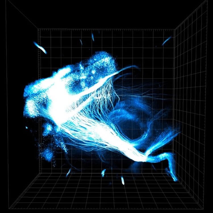
Image Source: This NeuroscienceNews.com image is credited to Li Ye and Karl Deisseroth.
Original Research: Abstract for “Wiring and molecular features of prefrontal ensembles representing distinct experiences” by Li Ye, William E. Allen, Kimberly R. Thompson, Qiyuan Tian, Brian Hsueh, Charu Ramakrishnan, Ai-Chi Wang, Joshua H. Jennings, Avishek Adhikari, Casey H. Halpern, Ilana B. Witten, Alison L. Barth, Liqun Luo, Jennifer A. McNab, and Karl Deisseroth in Science. Published online May 26 2016 doi:10.1016/j.cell.2016.05.010
[cbtabs][cbtab title=”MLA”]Stanford. “Neurons in the Prefrontal Cortex are Built to Respond to Reward or Aversion.” NeuroscienceNews. NeuroscienceNews, 27 May 2016.
<https://neurosciencenews.com/prefrontal-cortex-optogenetics-reward-aversion-4324/>.[/cbtab][cbtab title=”APA”]Stanford. (2016, May 27). Neurons in the Prefrontal Cortex are Built to Respond to Reward or Aversion. NeuroscienceNews. Retrieved May 27, 2016 from https://neurosciencenews.com/prefrontal-cortex-optogenetics-reward-aversion-4324/[/cbtab][cbtab title=”Chicago”]Stanford. “Neurons in the Prefrontal Cortex are Built to Respond to Reward or Aversion.” https://neurosciencenews.com/prefrontal-cortex-optogenetics-reward-aversion-4324/ (accessed May 27, 2016).[/cbtab][/cbtabs]
Abstract
Wiring and molecular features of prefrontal ensembles representing distinct experiences
Highlights
•Cohort-scale CLARITY is enabled for whole-brain activity/projection measurements
•mPFC cells active during distinct-valence experiences exhibit distinct projections
•Positive-valence experience preferentially recruits an NPAS4+ population in mPFC
•Recruiting diverse experience-defined mPFC ensembles drives distinct behaviors
Summary
A major challenge in understanding the cellular diversity of the brain has been linking activity during behavior with standard cellular typology. For example, it has not been possible to determine whether principal neurons in prefrontal cortex active during distinct experiences represent separable cell types, and it is not known whether these differentially active cells exert distinct causal influences on behavior. Here, we develop quantitative hydrogel-based technologies to connect activity in cells reporting on behavioral experience with measures for both brain-wide wiring and molecular phenotype. We find that positive and negative-valence experiences in prefrontal cortex are represented by cell populations that differ in their causal impact on behavior, long-range wiring, and gene expression profiles, with the major discriminant being expression of the adaptation-linked gene NPAS4. These findings illuminate cellular logic of prefrontal cortex information processing and natural adaptive behavior and may point the way to cell-type-specific understanding and treatment of disease-associated states.
“Wiring and molecular features of prefrontal ensembles representing distinct experiences” by Li Ye, William E. Allen, Kimberly R. Thompson, Qiyuan Tian, Brian Hsueh, Charu Ramakrishnan, Ai-Chi Wang, Joshua H. Jennings, Avishek Adhikari, Casey H. Halpern, Ilana B. Witten, Alison L. Barth, Liqun Luo, Jennifer A. McNab, and Karl Deisseroth in Science. Published online May 26 2016 doi:10.1016/j.cell.2016.05.010