Summary: Researchers report using primary targeted muscle reinnervation during amputation helps to reduce, and in some cases, prevent phantom limb and stump pain in patients.
Source: Ohio State University.
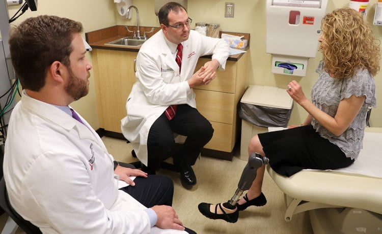
Doctors at The Ohio State University Wexner Medical Center and College of Medicine are pioneering the use of primary targeted muscle reinnervation (TMR) to prevent or reduce debilitating phantom limb and stump pain in amputees.
Losing a limb due to trauma, cancer or poor circulation can result in phantom limb and stump pain in upwards of 75 percent of amputees in the United States. Primary TMR – the rerouting of nerves cut during amputation into surrounding muscle – greatly reduces phantom limb and residual limb pain, as reported in recent publications by Dr. Ian Valerio, division chief of Burn, Wound and Trauma in Ohio State’s Department of Plastic and Reconstructive Surgery, and Dr. J. Byers Bowen, a former resident who is now in private practice. Their latest work featured in the January 2019 issue of Plastic and Reconstructive Surgery describes how to perform this technique in below-the-knee amputations.
TMR was first developed to allow amputees better control of upper limb prosthetics. Traditionally doctors perform the surgery months or years after the initial amputation. When surgeons discovered the procedure also improves certain causes of pain, they started using it to treat disorganized nerve endings called symptomatic neuromas and/or phantom limb pain.
In this paper, Valerio and Bowen provide a detailed description of TMR in below-the-knee amputees and document the benefits of primary TMR for preventing pain.
“This paper provides a blueprint for improving patient outcomes and quality of life following amputation,” said Dr. K. Craig Kent, dean of The Ohio State University College of Medicine.
Over the course of three years, the surgeons performed 22 TMR surgeries on below-the-knee amputees, 18 primary and four secondary. None of the patients has developed symptomatic neuromas and only 13 percent of patients who received primary TMR reported having pain six months later.
“A significant amount of pain in amputees is caused by disorganized nerve endings, i.e. symptomatic neuromas, in the residual limb. They form when nerves are severed and not addressed, thus they have nowhere to go,” Valerio said. “Attaching those cut nerve endings to motor nerves in a nearby muscle allows the body to re-establish its neural circuitry. This alleviates phantom and residual limb pain by giving those severed nerves somewhere to go and something to do.”
Valerio said patients who’ve had TMR significantly reduce or sometimes stop using narcotics and other nerve pain related medications, which can greatly improve their quality of life.

“TMR has been shown to reduce pain scores and multiple types of pain via a variety of validated pain surveys. These findings are the first to show that surgery can greatly reduce phantom and other types of limb pain directly,” Valerio said.
Bowen added that upper extremity amputees are better able to use and control their prosthetics in addition to their improved pain outcomes. He said, “TMR allows for more individual muscle unit firings through the patient’s thoughts. It provides for better intuitive control resulting in more refined functional movements and more degrees of motion by an advanced prosthetic.”
The researchers believe primary TMR is a reliable technique to prevent the development of disorganized nerve endings and to reduce phantom and other limb pain in all types of amputations. When done at the time of initial amputation, there is minimal health risk and recovery is similar to that of traditional amputation surgery.
Surgeons perform TMR routinely at Ohio State, with primary TMR as the standard of care for most orthopedic-based traumatic and oncologic amputations. Valerio lectures and trains surgeons around the world on the primary TMR technique in an effort to make it a global best practice.
Source: Serena Smith – Ohio State University
Publisher: Organized by NeuroscienceNews.com.
Image Source: NeuroscienceNews.com image is credited to Ohio State University.
Video Source: Video credited to Ohio State Wexner Medical Center.
Original Research: Abstract for “Targeted Muscle Reinnervation Technique in Below-Knee Amputation” by Bowen, J. Byers, M.D., M.S.; Ruter, Daniel, B.S.; Wee, Corinne, M.D.; West, Julie, M.S., P.A.-C.; Valerio, and Ian L., M.D., M.S., M.B.A. in Plastic and Reconstructive Surgery. Published December 31 2018.
doi:10.1097/PRS.0000000000005133
[cbtabs][cbtab title=”MLA”]Ohio State University”Rerouting Nerves During Amputation Reduces Phantom Limb Pain Before it Starts.” NeuroscienceNews. NeuroscienceNews, 31 December 2018.
<https://neurosciencenews.com/phantom-limb-pain-nerve-rerouting-10404/>.[/cbtab][cbtab title=”APA”]Ohio State University(2018, December 31). Rerouting Nerves During Amputation Reduces Phantom Limb Pain Before it Starts. NeuroscienceNews. Retrieved December 31, 2018 from https://neurosciencenews.com/phantom-limb-pain-nerve-rerouting-10404/[/cbtab][cbtab title=”Chicago”]Ohio State University”Rerouting Nerves During Amputation Reduces Phantom Limb Pain Before it Starts.” https://neurosciencenews.com/phantom-limb-pain-nerve-rerouting-10404/ (accessed December 31, 2018).[/cbtab][/cbtabs]
Abstract
Targeted Muscle Reinnervation Technique in Below-Knee Amputation
Cortical sensory neurons are characterized by selectivity to stimulation. This selectivity was originally viewed as a part of the fundamental “receptive field” characteristic of neurons. This view was later challenged by evidence that receptive fields are modulated by stimuli outside of the classical receptive field. Here, we show that even this modified view of selectivity needs revision. We measured spatial frequency selectivity of neurons in cortical area MT of alert monkeys and found that their selectivity strongly depends on luminance contrast, shifting to higher spatial frequencies as contrast increases. The changes of preferred spatial frequency are large at low temporal frequency, and they decrease monotonically as temporal frequency increases. That is, even interactions among basic stimulus dimensions of luminance contrast, spatial frequency, and temporal frequency strongly influence neuronal selectivity. This dynamic nature of neuronal selectivity is inconsistent with the notion of stimulus preference as a stable characteristic of cortical neurons.


