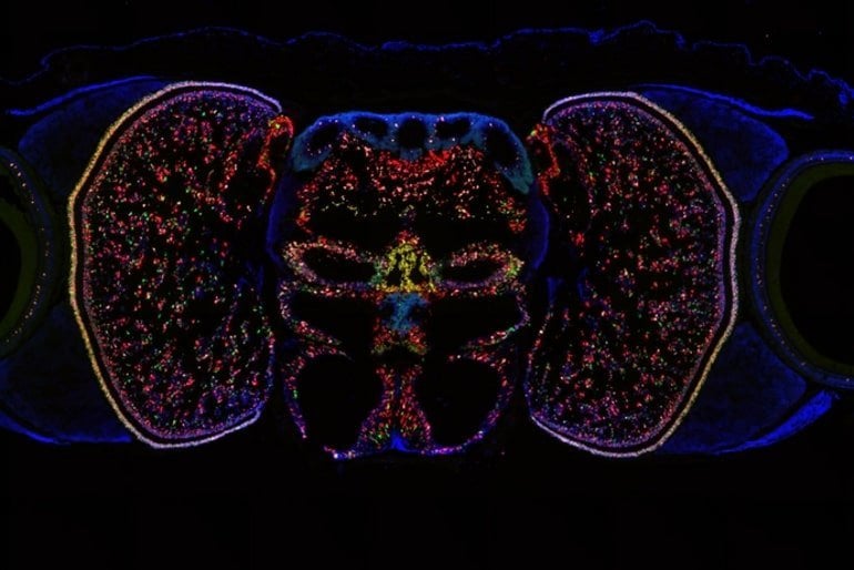
Summary: A new map of the octopus visual system classifies different types of neurons in a part of the brain dedicated to vision, shedding new light on the evolution of the brain and visual systems in a more broad sense.
Source: University of Oregon
It’s hard for the octopus to pick just one party trick. It swims via jet propulsion, shoots inky chemicals at its foes, and can change its skin within seconds to blend in with its surroundings.
A team of University of Oregon researchers is investigating yet another distinctive feature of this eight-armed marine animal: its outstanding visual capabilities.
In a new paper, they lay out a detailed map of the octopus’s visual system, classifying different types of neurons in a part of the brain devoted to vision. The map is a resource for other neuroscientists, giving details that could guide future experiments. And it could teach us something about the evolution of brains and visual systems more broadly, too.
The team reports their findings October 31 in Current Biology.
Cris Niell’s lab at the UO studies vision, mostly in mice. But a few years ago, postdoc Judit Pungor brought a new species to the lab — the California two-spot octopus.
While not traditionally used as a study subject in the lab, this cephalopod quickly captured the interest of UO neuroscientists. Unlike mice, which are not known for having good vision, “octopuses have an amazing visual system, and large fraction of their brain is dedicated to visual processing,” Niell said. “They have an eye that’s remarkably similar to the human eye, but after that, the brain is completely different.
The last common ancestor between octopuses and humans was 500 million years ago, and the species have since evolved in very different contexts. So scientists didn’t know whether the parallels in visual systems extended beyond the eyes, or whether the octopus was instead using completely different kinds of neurons and brain circuits to achieve similar results.
“Seeing how the octopus eye convergently evolved similarly to ours, it’s cool to think about how the octopus visual system could be a model for understanding brain complexity more generally,” said Mea Songco-Casey, a graduate student in Niell’s lab and the first author on the paper. “For example, are there fundamental cell types that are required for this very intelligent, complex brain?”
Here, the team used genetic techniques to identify different types of neurons in the octopus’s optic lobe, the part of the brain that’s devoted to vision.
They picked out six major classes of neurons, distinguished based on the chemical signals they send. Looking at the activity of certain genes in those neurons then revealed further subtypes, providing clues to more specific roles.
In some cases, the researchers pinpointed particular groups of neurons in distinctive spatial arrangements — for instance, a ring of neurons around the optic lobe that all signal using a molecule called octopamine. Fruit flies use this molecule, which is similar to adrenaline, to increase visual processing when the fly is active. So it could perhaps have a similar role in octopuses.
“Now that we know there’s this very specific cell type, we can start to go in and figure out what it does,” Niell said.
About a third of the neurons in the data didn’t quite look fully developed. The octopus brain keeps growing and adding new neurons over the animal’s lifespan. These immature neurons, not yet integrated into brain circuits, were a sign of the brain in the process of expanding!

However, the map didn’t reveal sets of neurons that clearly transferred over from humans or other mammalian brains, as the researchers thought it might.
“At the obvious level, the neurons don’t map onto each other—they’re using different neurotransmitters,” Niell said. “But maybe they’re doing the same kinds of computations, just in a different way.”
Digging deeper will also require getting a better handle on cephalopod genetics. Because the octopus hasn’t traditionally been used as a lab animal, many of the tools that are used for precise genetic manipulation in fruit flies or mice don’t yet exist for the octopus, said Gabby Coffing, a graduate student in Andrew Kern’s lab who worked on the study.
“There are a lot of genes where we have no idea what their function is, because we haven’t sequenced the genomes of a lot of cephalopods,” Pungor said. Without genetic data from related species as a point of comparison, it’s harder to deduce the function of particular neurons.
Niell’s team is up for the challenge. They’re now working to map the octopus brain beyond the optic lobe, seeing how some of the genes they focused on in this study show up elsewhere in the brain. They are also recording from neurons in the optic lobe, to determine how they process the visual scene.
In time, their research might make these mysterious marine animals a little less murky — and shine a little light on our own evolution, too.
About this brain mapping and visual neuroscience research news
Author: Laurel Hamers
Source: University of Oregon
Contact: Laurel Hamers – University of Oregon
Image: The image is credited to Niell Lab
Original Research: Open access.
“Cell types and molecular architecture of the Octopus bimaculoides visual system” by Cris Niell et al. Current Biology
Abstract
Cell types and molecular architecture of the Octopus bimaculoides visual system
Highlights
- scRNA-seq and FISH identified molecular cell types in the octopus visual system
- Cell types defined by functional and developmental markers show sublayer organization
- Immature neurons form transcriptional subgroups that correspond to mature cell types
- This atlas is a basis for studying visual function and development in cephalopods
Summary
Cephalopods have a remarkable visual system, with a camera-type eye and high acuity vision that they use for a wide range of sophisticated visually driven behaviors.
However, the cephalopod brain is organized dramatically differently from that of vertebrates and invertebrates, and beyond neuroanatomical descriptions, little is known regarding the cell types and molecular determinants of their visual system organization.
Here, we present a comprehensive single-cell molecular atlas of the octopus optic lobe, which is the primary visual processing structure in the cephalopod brain.
We combined single-cell RNA sequencing with RNA fluorescence in situ hybridization to both identify putative molecular cell types and determine their anatomical and spatial organization within the optic lobe.
Our results reveal six major neuronal cell classes identified by neurotransmitter/neuropeptide usage, in addition to non-neuronal and immature neuronal populations.
We find that additional markers divide these neuronal classes into subtypes with distinct anatomical localizations, revealing further diversity and a detailed laminar organization within the optic lobe.
We also delineate the immature neurons within this continuously growing tissue into subtypes defined by evolutionarily conserved developmental genes as well as novel cephalopod- and octopus-specific genes.
Together, these findings outline the organizational logic of the octopus visual system, based on functional determinants, laminar identity, and developmental markers/pathways.
The resulting atlas presented here details the “parts list” for neural circuits used for vision in octopus, providing a platform for investigations into the development and function of the octopus visual system as well as the evolution of visual processing.


