

Summary:
Biomedical engineers at Penn State have solved a major hurdle in neuroprosthetics and sensory rehabilitation: sensory habituation, where the brain gradually stops perceiving continuous artificial electrical stimulation. In a paper published in IEEE Transactions on Medical Imaging, researchers led by Dr. Xiaogang Hu monitored cortical activity using functional near-infrared spectroscopy (fNIRS) during transcutaneous nerve stimulation (TNS).
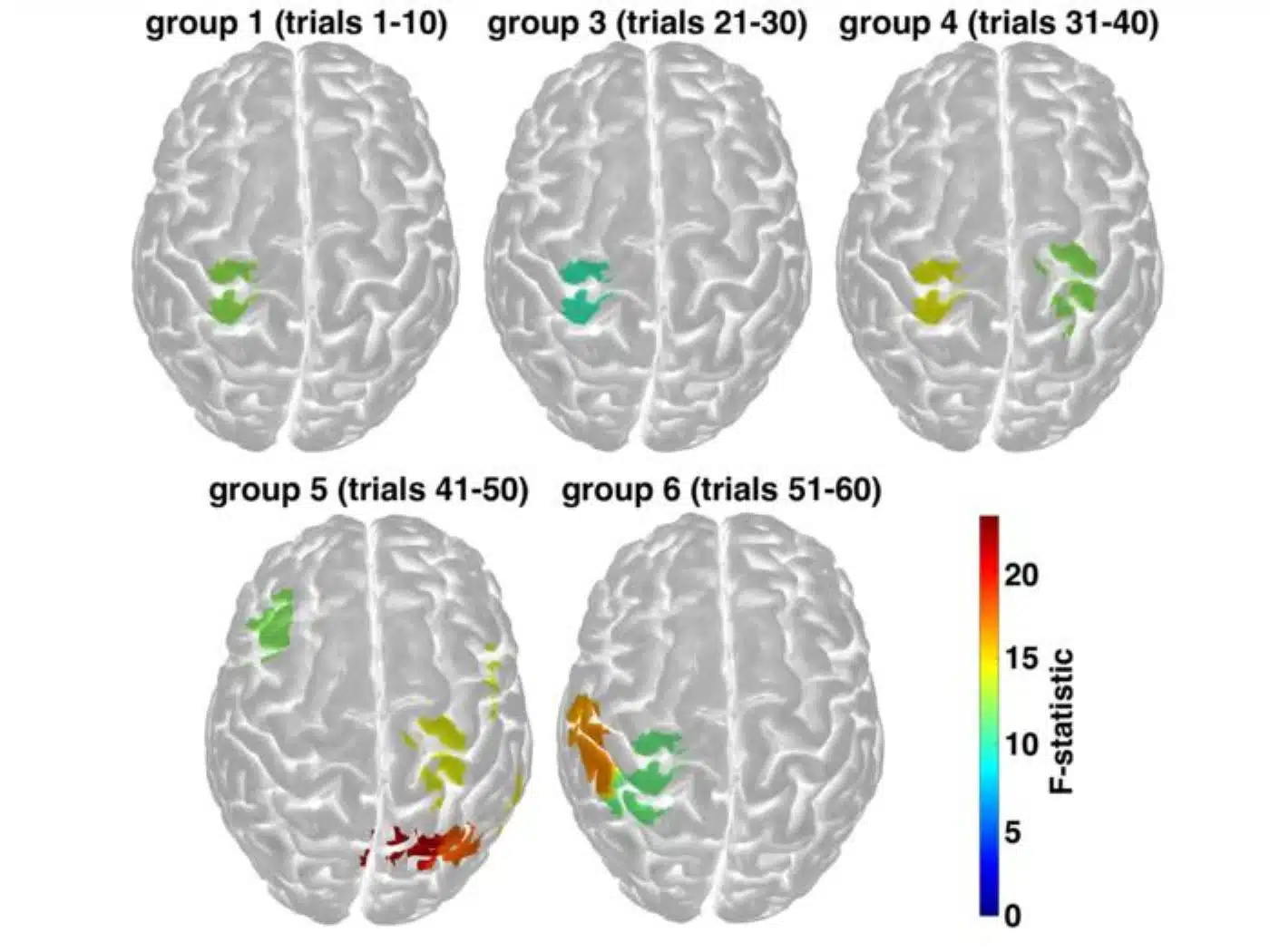
By delivering 5-second electrical pulse bursts paired with 40-second recovery intervals, the team maintained a stable, unattenuated sensation of touch over 45 minutes across 60 trials. Neuroimaging revealed that while perception remained completely constant, the brain actively distributed tactile processing beyond the primary somatosensory cortex to wider neural networks.
Key Facts:
- Overcoming Sensory Fading: Habituation, the brain’s natural tendency to mute repetitive sensory inputs, routinely causes artificial tactile stimulation to fade over time. The new protocol eliminated this perceptual decline over a 45-minute testing window.
- The 5-to-40 Optimal Ratio: Testing various pulse durations and recovery windows identified an optimal parameters “sweet spot”: 5 seconds of mild transcutaneous electrical nerve stimulation followed by 40 seconds of rest.
- Cortical Network Migration: Non-invasive fNIRS neuroimaging revealed that even though participants perceived no change in sensation intensity, active cortical regions migrated over time, moving from the focal somatosensory cortex to recruit broader, distributed neural processing networks.
Source: Penn State
Loss of tactile sensation in the hands, whether resulting from stroke, peripheral nerve injury, spinal trauma, or limb amputation, fundamentally impairs everyday activities. Basic tasks such as holding a fragile ceramic mug, grasping utensils, writing, or typing become daily battles when real-time sensory feedback is absent.
While bioengineers have developed transcutaneous nerve stimulation (TNS) and invasive neural implants capable of delivering artificial electrical pulses to mimic the sensation of touch, these technologies face a universal physiological barrier: sensory habituation.
Over extended periods, the central nervous system adapts to repetitive, artificial electrical stimulation. As neural circuits adjust to constant inputs, perceived intensity weakens and eventually fades away entirely. For an amputee using a motorized prosthetic hand, this decay is catastrophic: without steady tactile feedback, an individual cannot regulate grip force, leading them to either crush delicate objects or drop them unexpectedly.
Now, a team of researchers led by Dr. Xiaogang Hu, the Dorothy Foehr Huck and J. Lloyd Huck Chair in Neurorehabilitation and professor of mechanical engineering at Penn State, has uncovered both the neural dynamics of habituation and a stimulation protocol to bypass it.
Published in IEEE Transactions on Medical Imaging, the study demonstrates that finely calibrated stimulation-and-rest intervals can preserve stable, long-term tactile sensations while triggering adaptive redistribution across the brain’s cerebral networks.
Pinpointing the Stimulation “Sweet Spot”
“Repetitive stimulation — regardless of whether you engage stimulation through the nerves, skin or directly targeting a part of the brain — will be perceived as declining or weakening over time,” explained Dr. Hu, who holds joint appointments in Penn State’s Departments of Kinesiology and Physical Medicine and Rehabilitation. “With this work, we are trying to avoid or minimize that habituation, enabling a more stable sense of touch over a longer timeframe.”
To determine how electrical parameters influence neural adaptation, the researchers systematically tested varying electrical pulse durations and resting intervals across healthy volunteers.
Electrodes were placed over peripheral nerves in the forearm to deliver mild electrical pulses designed to feel like a gentle tingling sensation. Using trial-by-trial psychophysical scoring, the team determined that a specific combination, 5 seconds of electrical stimulation followed by a 40-second recovery interval, prevented sensory decay.
When evaluated across an extended 45-minute continuous trial consisting of 60 consecutive stimulation cycles, participants reported zero habituation, retaining a crisp, consistent tactile sensation from the first pulse to the last.
“Picture someone is holding a cup of water with their prosthetic — the sensation of touch is critical for them to maintain an appropriate grip force,” Hu noted. “We don’t want them to crush the cup with too much force or for them to lose their sensation of touch over time and drop the cup; we need to ensure that the feeling is stable.”
Peering Beneath the Skull with Portable fNIRS
To explore how the brain maintains stable perception during prolonged stimulation, the investigators deployed functional near-infrared spectroscopy (fNIRS).
Unlike functional magnetic resonance imaging (fMRI), which requires patients to lie immobilized inside a massive, noisy scanner bore, fNIRS employs a flexible cap equipped with lightweight optical optodes. These emitters project harmless beams of near-infrared light through the scalp and skull into the superficial cerebral cortex. By measuring how light reflects back to paired photodetectors, the system quantifies local shifts in oxygenated and deoxygenated hemoglobin, serving as a high-fidelity proxy for localized neuronal metabolism and synaptic activity.
Targeting the primary somatosensory cortex (the cerebral hub responsible for mapping bodily touch), fNIRS captured real-time cortical dynamics while participants sat upright and received TNS.
The optical scans uncovered an unexpected neurofunctional divergence: while participants reported that the physical sensation felt identical across all 45 minutes, their underlying brain activation patterns were constantly shifting.
“Participants report that the sensation feels the same, but the active regions in their brain actually change over time, with changing intensity of activation and spatial distribution,” said first author Dr. Susan Coltman, assistant professor at the University of Guelph, who conducted the study as a postdoctoral researcher in Hu’s lab. “The signals start out in the somatosensory cortex, but over time, they move out beyond that origin area to different regions of the brain, reflecting progressive recruitment of distributed brain regions to process information during prolonged sensory stimulation.”
Rewiring the Future of Neuroprosthetics
Rather than a static, local response, sensory preservation appears to depend on the brain dynamically recruiting distributed associative and secondary sensory networks to sustain conscious perception.
These findings carry major implications for clinical neurorehabilitation. Individuals recovering from hemispheric stroke or living with upper-limb amputations already experience extensive cortical reorganization. Understanding how the healthy brain balances stimulation timing and network recruitment provides a quantitative template for programming next-generation bionic prostheses and wearable rehabilitation sleeves.
The research team plans to extend these optimized stimulation paradigms into patient populations with focal stroke lesions and limb loss, aiming to calibrate personalized electrical intervals that accommodate damaged, rewired neural pathways.
By striking the biological balance between stimulation and recovery, engineers are moving closer to artificial limbs that don’t just move naturally, but feel permanently real.
Questions and Answers
What is sensory habituation in nerve stimulation?
Sensory habituation occurs when the nervous system becomes accustomed to continuous, repetitive artificial electrical stimulation, causing the perceived sensation of touch to gradually weaken and fade away over time.
How did the Penn State team prevent habituation during nerve stimulation?
The researchers identified an optimized timing ratio: applying 5 seconds of mild electrical stimulation followed by a 40-second rest period. This cycle allowed peripheral nerves and central circuits to recover, sustaining a completely stable sense of touch over 45 minutes and 60 cycles.
How did brain activity change during the experiment?
Although participants felt the exact same level of sensation throughout the 45-minute window, fNIRS imaging showed that active brain regions shifted over time. Brain activity originated in the primary somatosensory cortex and then progressively migrated outward, recruiting wider distributed brain networks to sustain sensory processing.
Funding & Support: Supported by the National Institute on Disability, Independent Living, and Rehabilitation Research (NIDILRR) and the U.S. National Science Foundation’s Division of Information and Intelligent Systems (grants IIS-2330862 and IIS-2319139).
Editorial Notes:
- This article was edited by a Neuroscience News editor.
- Journal paper reviewed in full.
- Additional context added by our staff.
About this neuroscience and neurotech Research:
- Media Contact: Ty Tkacik
- Source: Penn State
- Image Credit: Image credited to Xiaogang Hu
- Original Research is Open Access: IEEE Transactions on Medical Imaging (Nov 11, 2026). “fNIRS Characterization of Temporal Potentiation During Optimized Nerve Stimulation.” Authors: Susan Coltman, William B. Riggs, Androu Abdalmalak, and Xiaogang Hu.
- DOI: 10.102110.1109/TMI.2026.3738278
Abstract
fNIRS Characterization of Temporal Potentiation During Optimized Nerve Stimulation
Restoring stable somatosensory feedback through transcutaneous nerve stimulation (TNS) represents a significant challenge in neural engineering; however, the cortical dynamics underlying sustained stimulation are not yet well characterized.
This study utilizes a multi-method functional near-infrared spectroscopy framework to quantify the temporal evolution of cortical engagement during temporally structured TNS. It was hypothesized that stimulation parameters fundamentally constrain the spatiotemporal profile of the hemodynamic response function.
A General Linear Model with temporal and dispersion derivatives was used to analyze hemodynamic responses in 10 participants across three phases: validation, duration optimization (2, 5, or 10 seconds), and sustained engagement (45 minutes). During the optimization phase, 5-second stimulation produced the highest canonical amplitude with optimal dispersion characteristics, while 10-second trains resulted in significant temporal delays.
During the sustained phase, parametric modeling indicated no linear decay in cortical response. Instead, the cortex demonstrated temporal potentiation, characterized by a transition from sparse early-phase activation to widespread late-phase recruitment across the sensorimotor network. The left posterior parietal cortex (BA5) served as a continuous integration hub, whereas primary somatosensory regions exhibited dynamic late-onset engagement.
These findings characterize a nonlinear temporal potentiation pattern, demonstrating that the selected stimulation/rest timing regime can sustain cortical engagement and facilitate delayed network expansion. The observed intersubject heterogeneity highlights the necessity for adaptive, closed-loop TNS controllers guided by real-time hemodynamic biomarkers.


