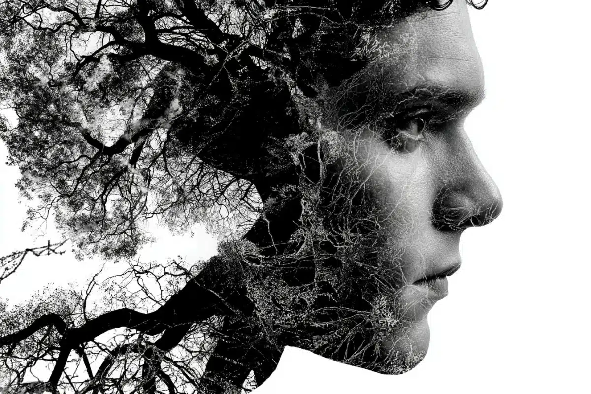
Scientists have discovered differences in the brain structure of ballet dancers that may help them avoid feeling dizzy when they perform pirouettes.
The research suggests that years of training can enable dancers to suppress signals from the balance organs in the inner ear.
The findings, published in the journal Cerebral Cortex, could help to improve treatment for patients with chronic dizziness. Around one in four people experience this condition at some time in their lives.
Normally, the feeling of dizziness stems from the vestibular organs in the inner ear. These fluid-filled chambers sense rotation of the head through tiny hairs that sense the fluid moving. After turning around rapidly, the fluid continues to move, which can make you feel like you’re still spinning.
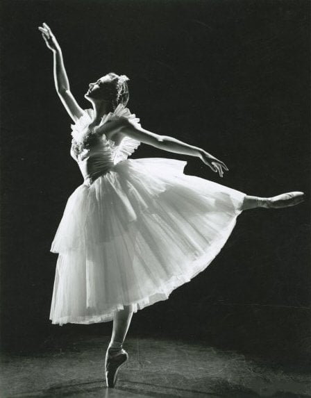
Ballet dancers can perform multiple pirouettes with little or no feeling of dizziness. The findings show that this feat isn’t just down to spotting, a technique dancers use that involves rapidly moving the head to fix their gaze on the same spot as much as possible.
Researchers at Imperial College London recruited 29 female ballet dancers and, as a comparison group, 20 female rowers whose age and fitness levels matched the dancers’.
The volunteers were spun around in a chair in a dark room. They were asked to turn a handle in time with how quickly they felt like they were still spinning after they had stopped. The researchers also measured eye reflexes triggered by input from the vestibular organs. Later, they examined the participants’ brain structure with MRI scans.
In dancers, both the eye reflexes and their perception of spinning lasted a shorter time than in the rowers.
Dr Barry Seemungal, from the Department of Medicine at Imperial, said: “Dizziness, which is the feeling that we are moving when in fact we are still, is a common problem. I see a lot of patients who have suffered from dizziness for a long time. Ballet dancers seem to be able to train themselves not to get dizzy, so we wondered whether we could use the same principles to help our patients.”
The brain scans revealed differences between the groups in two parts of the brain: an area in the cerebellum where sensory input from the vestibular organs is processed and in the cerebral cortex, which is responsible for the perception of dizziness.
The area in the cerebellum was smaller in dancers. Dr Seemungal thinks this is because dancers would be better off not using their vestibular systems, relying instead on highly co-ordinated pre-programmed movements.
“It’s not useful for a ballet dancer to feel dizzy or off balance. Their brains adapt over years of training to suppress that input. Consequently, the signal going to the brain areas responsible for perception of dizziness in the cerebral cortex is reduced, making dancers resistant to feeling dizzy.
“If we can target that same brain area or monitor it in patients with chronic dizziness, we can begin to understand how to treat them better.”
Another finding in the study may be important for how chronic dizzy patients are tested in the clinic. In the control group, the perception of spinning closely matched the eye reflexes triggered by vestibular signals, but in dancers, the two were uncoupled.
“This shows that the sensation of spinning is separate from the reflexes that make your eyes move back and forth,” Dr Seemungal said. “In many clinics, it’s common to only measure the reflexes, meaning that when these tests come back normal the patient is told that there is nothing wrong. But that’s only half the story. You need to look at tests that assess both reflex and sensation.”
Notes about this neuroscience and neurobiology research
The research was funded by a Health Foundation / Academy of Medical Sciences Fellowship and the Medical Research Council.
Written by Sam Wong
Contact: Sam Wong – Imperial College London
Source: Imperial College London press release
Image Source: The photograph of Jocelyn Vollmar is credited to the National Library of Australia and is in the public domain.
Original Research: Full open access research for “The Neuroanatomical Correlates of Training-Related Perceptuo-Reflex Uncoupling in Dancers” by Yuliya Nigmatullina, Peter J. Hellyer, Parashkev Nachev, David J. Sharp and Barry M. Seemungal⇑ in Cerebral Cortex. Published online September 27 2013 doi:10.1093/cercor/bht266