

In the middle of the human brain there is a tiny structure shaped like an elongated donut that plays a crucial role in managing how the body functions. Measuring just 10 millimeters in length and six millimeters in diameter, the hollow structure is involved in a complex array of behavioral, cognitive, and affective phenomena, such as the fight or flight response, pain regulation, and even sexual activity, according to Northeastern senior research scientist Ajay Satpute.
With a name longer than the structure itself, the “midbrain periaqueductal gray region,” or PAG, is extraordinarily difficult to investigate in humans because of its size and intricate structure, he said.
In research published online this week in the journal Proceedings of the National Academy of Science, Satpute and his colleagues at Northeastern’s Interdisciplinary Affective Science Laboratory explain how they hurdled these challenges by using state-of-the art imaging to capture this complex neural activity. The research could ultimately help scientists explore the grounds of human emotion like never before.

“The PAG’s functional properties occur at such small spatial scales that we need to capture its activity at very high resolution in order to understand it,” he explained.
Until recently, neuroimaging studies have been carried out on functional magnetic resonance imaging, or fMRI, instruments containing magnets of up to three Teslas, a measure of magnetic field strength. These instruments provide critical data for understanding how the brain’s different areas respond to different stimuli, but when those areas become sufficiently small and complicated, their resolution falls short.
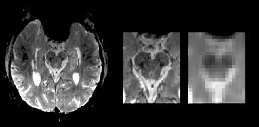
In the case of the tiny PAG, this problem is paramount because the PAG wraps around a hollow core, or “aqueduct,” containing cerebrospinal fluid, Satpute said. Traditional fMRI instruments cannot distinguish neural activity occurring in the PAG from that occurring in the CS fluid. Even more difficult is identifying where within the PAG itself specific responses originate.
In collaboration with researchers at the Massachusetts General Hospital in Boston, Satpute and his colleagues used a high-tech fMRI instrument that contains a seven-Tesla magnet. The force of the instrument is so strong (albeit harmless) that one can feel its pull when simply walking by. Coupled with painstaking manual data analyses, Satpute was able to resolve activity in sub-regions of the PAG with more precision than ever before.
With their method in hand, the research team showed 11 human research subjects images of burn victims, gory injuries, and other content related to threat, harm, and loss while keeping tabs on the PAG’s activity. Researchers also showed the subjects neutral images such and then compared results between the two scenarios.
The proof-of-concept study showed emotion-related activity concentrated in particular areas of the PAG. While similar results have been demonstrated in animal models, nothing like it had previously been shown in human brains.
Using this methodology, the researchers said they would not only gain a better understanding of the PAG but also be able to investigate a range of brain-related research questions beyond this particular structure.
Seven-Tesla brain imaging provides an unprecedented view of regions like the PAG while they respond to stimuli, said Lisa Feldman Barrett, director of the Interdisciplinary Affective Science Laboratory. “Studies like this are a critical step forward in bridging human and nonhuman animal studies of emotion, because they offer a level of resolution in human brains that was previously possible only in studies of non-human animal,” she said.
Notes about this neuroimaging research
Written by Angela Herring
Contact: Angela Herring – Northeastern University
Source: Northeastern University press release
Image Source: The image is credited to Ajay Satpute, Northeastern University and is adapted from the press release.
Original Research: Abstract for “Identification of discrete functional subregions of the human periaqueductal gray” by Ajay B. Satpute, Tor D. Wager, Julien Cohen-Adad, Marta Bianciardi, Ji-Kyung Choi, Jason T. Buhle, Lawrence L. Wald, and Lisa Feldman Barrett in PNAS. Published online September 30 2013 doi:10.1073/pnas.1306095110


