Summary: A newly developed headset capable of brain mapping may help provide new insight into developmental disorders, including autism and cerebral palsy.
Source: UCL
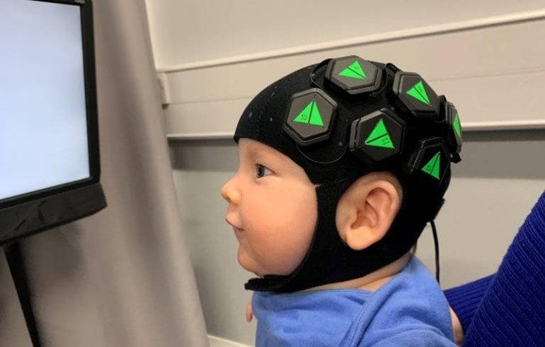
A team led by UCL researchers has demonstrated a new form of wearable, baby-friendly brain mapping technology that has important implications for understanding developmental conditions such as autism spectrum disorder and cerebral palsy.
The technology uses harmless levels of red and near-infrared light delivered via a wearable cap to generate detailed 3D images of babies’ brain activity. It means doctors and neuroscientists can image a baby’s brain without needing MRI, which is expensive and difficult to apply while babies are awake.
It also means researchers can study the infant brain in almost any environment, including in the home where babies and parents naturally interact.
In the paper, published in NeuroImage, academics and engineers from UCL, Cambridge University, the Rosie Hospital in Cambridge and London-based start-up company Gowerlabs Ltd demonstrate the technique, known as high-density diffuse optical tomography (HD-DOT), in six-month old infants. The wearable cap employs hundreds of LEDs and optical detectors arranged in a dense network over the scalp to map changes in oxygenation in the brain.
These changes in oxygenation show the areas of the brain that are busy processing information, meaning the team was were able to create high-quality, 3D images of baby brain activity for the first time outside an MRI scanner.
Project lead Dr Rob Cooper (UCL Medical Physics & Biomedical Engineering) explained: “There is a lot we still don’t know about how the brain develops, and a big part of the problem is that studying the infant brain is really difficult with traditional scanners.
“As any parent knows, 6-month old babies are very active; they move around all the time and are easily distracted. Using a technique like MRI, the subject has to remain completely still, which is almost impossible with babies unless they are asleep or sedated.”
Elisabetta Maria Frijia (UCL Medical Physics & Biomedical Engineering), who carried out much of the work demonstrating the technology, added: “The approach we have demonstrated is safe, silent and wearable, and can produce images of brain function with better spatial resolution than any other comparable technology.
“Our hope is that this new generation of technologies will allow researchers from a whole range of fields to learn more about how the healthy infant brain develops and establish new ways of diagnosing, monitoring and ultimately treating neurological conditions like autism and cerebral palsy.”
The technology was developed by UCL spinout company Gowerlabs Ltd., whilst the work demonstrating its capabilities was led by Dr Cooper, with support from an EPSRC Early Career Fellowship.
About this neurotech research news
Source: UCL
Contact: Kate Corry – UCL
Image: The image is credited to Robert Cooper
Original Research: Closed access.
“Functional imaging of the developing brain with wearable high-density diffuse optical tomography: A new benchmark for infant neuroimaging outside the scanner environment” by Rob Cooper et al. NeuroImage
Abstract
Functional imaging of the developing brain with wearable high-density diffuse optical tomography: A new benchmark for infant neuroimaging outside the scanner environment
Studies of cortical function in the awake infant are extremely challenging to undertake with traditional neuroimaging approaches. Partly in response to this challenge, functional near-infrared spectroscopy (fNIRS) has become increasingly common in developmental neuroscience, but has significant limitations including resolution, spatial specificity and ergonomics. In adults, high-density arrays of near-infrared sources and detectors have recently been shown to yield dramatic improvements in spatial resolution and specificity when compared to typical fNIRS approaches. However, most existing fNIRS devices only permit the acquisition of ~20–100 sparsely distributed fNIRS channels, and increasing the number of optodes presents significant mechanical challenges, particularly for infant applications. A new generation of wearable, modular, high-density diffuse optical tomography (HD-DOT) technologies has recently emerged that overcomes many of the limitations of traditional, fibre-based and low-density fNIRS measurements. Driven by the development of this new technology, we have undertaken the first study of the infant brain using wearable HD-DOT. Using a well-established social stimulus paradigm, and combining this new imaging technology with advances in cap design and spatial registration, we show that it is now possible to obtain high-quality, functional images of the infant brain with minimal constraints on either the environment or on the infant participants. Our results are consistent with prior low-density fNIRS measures based on similar paradigms, but demonstrate superior spatial localization, improved depth specificity, higher SNR and a dramatic improvement in the consistency of the responses across participants. Our data retention rates also demonstrate that this new generation of wearable technology is well tolerated by the infant population.