Summary:
Neuroscientists discovered a previously hidden full-body map within the secondary motor cortex (M2), a brain region long thought to lack somatotopic organization. The study used high-speed wide-field optical imaging to analyze resting-state brain activity in mice, revealing synchronized sub-regions linking M2 to the sensory cortex that activate during complex, multi-limb actions.
Key Facts:
- A Hidden Somatotopic Map: While the primary motor cortex (M1) is well known for its orderly body map, researchers uncovered a distinct, previously overlooked body map embedded within the intrinsic resting-state neural chatter of the secondary motor cortex (M2).
- Synchronized Sensory-Motor Wiring: High-speed optical imaging and anatomical tracing confirmed that specific sub-regions within M2 are structurally wired to, and fire in synchrony with, corresponding body representations in the sensory cortex.
- Orchestrating Multi-Part Actions: Unlike M1, which governs isolated single-limb movements, M2 activates intensely during complex, coordinated behaviors requiring multiple body parts simultaneously, such as bringing food to the mouth.
Source: Columbia University
Coordinating the simultaneous motion of an arm, hand, head, and lips to sip a spoonful of soup feels effortless to the human brain, yet executing such fluid multi-part actions remains one of the most formidable challenges in robotics and computational neuroscience.
For decades, neuroscientists have understood that the primary motor cortex (M1) contains an orderly somatotopic “body map”, a spatial arrangement in which distinct cortical zones control individual muscles and body parts, such as a single finger or toe.
However, direct electrical stimulation of the neighboring secondary motor cortex (M2) yielded ambiguous results, leading researchers to conclude that M2 lacked a structured body map and instead operated purely as an abstract motor planning center.
Now, in a study published in Nature Communications, researchers led by Dr. Weihao Xu and Dr. Elizabeth Hillman at Columbia’s Mortimer B. Zuckerman Mind Brain Behavior Institute have overturned that assumption.
Using advanced wide-field optical neuroimaging, the team discovered a complete, previously hidden body map embedded within the resting-state neural activity of M2, revealing how the brain integrates sensory feedback to orchestrate complex, multi-limb movements.
Uncovering a Map Hidden in Resting Chatter
Studying motor control in mice poses a unique experimental hurdle because rodents rarely move a single body part in isolation. Rather than relying solely on artificial electrical stimulation or isolated movement tasks, Dr. Xu and colleagues utilized high-speed wide-field optical imaging techniques pioneered in the Hillman laboratory to capture cortex-wide neural dynamics in real time.
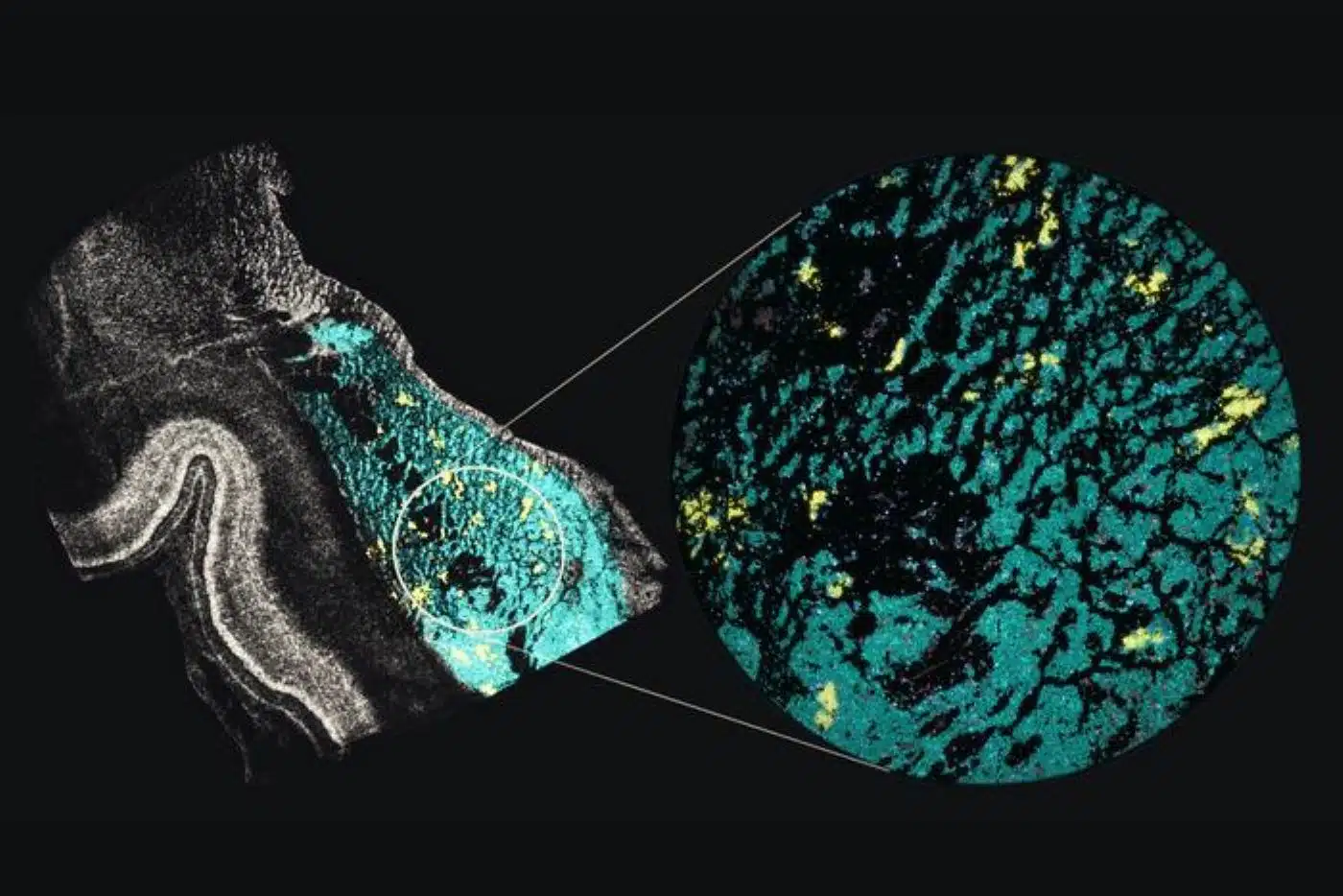
Unexpectedly, the critical breakthrough emerged not while the mice were actively moving, but while they were completely at rest. By analyzing spontaneous, seemingly random neural fluctuations across the resting cortex, the investigators spotted localized patches within M2 whose firing patterns synchronized precisely with established body-part sub-regions in the sensory cortex.
“We put these small spots together, and found that they formed a body map hidden in the intrinsic chatter of M2, a part of the brain that wasn’t supposed to have a body map,” said lead author Dr. Weihao Xu, who conducted the research as a doctoral student and postdoctoral scientist in Hillman’s lab.
To ensure these signals were not bleeding over from neighboring M1, Xu spent three years validating the finding using two-color wide-field optical mapping. By optically separating corticospinal projection neurons (which concentrate heavily in M1 and send direct commands down the spinal cord) from surrounding cortical populations, the team confirmed that the newly mapped zones resided squarely within M2.
Anatomical Wiring and Multi-Limb Coordination
Anatomical circuit tracing further confirmed that each sub-region of the newfound M2 body map maintained direct structural wiring with its corresponding counterpart in the sensory cortex.
When the researchers monitored these exact M2 zones during active behaviors, the functional purpose of this hidden map became clear. When a mouse performed a coordinated, multi-part action, such as raising both forepaws toward its mouth while simultaneously licking them, the corresponding forepaw, mouth, and tongue zones within the M2 map lit up together in intense, coordinated bursts.
“All of these features of M2 explain why understanding the activity patterns of M2 has been so challenging,” said corresponding author Dr. Elizabeth Hillman, now chair of the Department of Imaging Sciences at St. Jude Children’s Research Hospital. “Confusingly, M2 chatters when the body is still, and cares more about complicated movements than simple ones.”
Implications for Stroke Recovery and Robotics
By revealing how M2 bridges sensory maps with multi-limb motor execution, the findings offer a foundational biological blueprint for understanding how the brain plans and updates complex actions from moment to moment.
“This newfound map could help researchers understand key details about the dextrous movements that can be lost after brain injuries such as strokes,” Xu noted, adding that decoding how resting-state cortical chatter reflects underlying structural wiring opens new avenues for assessing circuit integrity after neurological trauma.
Beyond clinical neurorehabilitation and movement disorders, deciphering how biological brains effortlessly synchronize dozens of joints and sensory streams provides an architectural template for engineers designing next-generation, dexterous autonomous robots.
Questions and Answers
How does the secondary motor cortex (M2) differ from the primary motor cortex (M1)?
The primary motor cortex (M1) sends direct signals down the spinal cord to control simple, isolated movements of individual body parts. In contrast, the secondary motor cortex (M2) specializes in orchestrating complex, coordinated movements that require multiple body parts to work together simultaneously, such as bringing a cup to your lips.
How did researchers discover the hidden body map in M2?
Instead of relying on traditional electrical stimulation, researchers used high-speed wide-field optical imaging to watch brain activity while mice were resting. They discovered small patches in M2 whose spontaneous resting activity synchronized with known body-part regions in the sensory cortex, forming a complete, previously undetected map.
Why is this discovery important for stroke recovery and robotics?
Understanding how M2 maps and coordinates multi-limb movements helps neuroscientists pinpoint why complex dexterity is lost after strokes or brain injuries, potentially guiding targeted neurorehabilitation. It also gives robotics engineers a biological blueprint for programming fluid, multi-joint coordination.
Funding: Funding for this work was provided by National Institute of Neurological Disorders and Stroke BRAIN Initiative grants U19NS104649, R01 NS076628, R01 NS063226 and UM1NS132358 National Institute of Mental Health BRAIN Initiative grants RF1 MH114276 and RF1MH128969, the Simons Collaboration on the Global Brain grant 542991 and Foundation Leducq grant 22CVD01, and the American Lebanese Syrian Associated Charities (ALSAC), the fundraising and awareness organization of St. Jude.
Editorial Notes:
- This article was edited by a Neuroscience News editor.
- Journal paper reviewed in full.
- Additional context added by our staff.
About this neuroscience Research:
- Media Contact: Press Team
- Source: Zuckerman Institute
- Image Credit: Image credited to Neuroscience News
- Original Research is Open Access: Nature Communications (Nov 13, 2026). “Intrinsic Neural Dynamics Reveal a Body-Part-Specific Topography in Mouse Secondary Motor Cortex.” Authors: Weihao Xu, Alexis J. Yagielski, David N. Thibodeaux, Lina Marcela Carmona, Kosisochukwu Ugorji, Somayeh Shahsavarani, Sharon H. Kim, Fatema Lodgher, Hanzhi T. Zhao, Elizabeth A. Malan, Lu Xu, Caitlin Freeman, Anupam Bisht, Kaitlyn Swayze, Morgan Cambareri, Wenze Li & Elizabeth M. C. Hillman.
- DOI: 10.1038/s41467-026-78900-3
Abstract
Intrinsic Neural Dynamics Reveal a Body-Part-Specific Topography in Mouse Secondary Motor Cortex
The mammalian sensory cortex (S1) has a well-characterized body part-specific topography. However, most motor cortex atlases to date are only coarsely defined.
Here, we demonstrate that optical mapping of neuronal activity in awake mice reveals a distinct, body-part specific organization of mouse secondary motor cortex (M2).
This M2 topography was identified by recognizing that intrinsic neuronal fluctuations are selectively synchronized between homologous sub-regions of S1 and M2. These fluctuations were strongest during rest and quenched during movement, and were not prominent in corticospinal neurons (CSNs), indicating that they do not represent motor output.
Functional characterization of M2 sub-regions during spontaneous movements confirms body-part specificity, and stronger engagement during multi-effector coordinated actions. Structural connectivity atlas tracings from S1 regions confirm projections to homologous M2 sub-regions.
These results reveal an important underlying topography within M2, and demonstrate intrinsic neuronal dynamics as a window into the functional architecture of the motor cortex.