

Summary: A new study reveals how neurons implicated in the perception of motion are formed in the brains of fruit flies.
Source: NYU.
A team of biologists has deciphered how neurons used in the perception of motion form in the brain of a fly —a finding that illustrates how complex neuronal circuits are constructed from simple developmental rules.
The discovery, which appears in the journal Cell, offers new pathways for understanding the fundamental processes by which circuits in the brain form to process visual information.
“Understanding how neuronal identities are specified and how neural circuits are built during development leaves us in a better position to understand the onset of neurological disorders,” explains lead author Filipe Pinto-Teixeira, a post-doctoral fellow at New York University’s Department of Biology and NYU Abu Dhabi’s Center for Genomics and Systems Biology. “Specifically, we can fine tune our understanding of the generation of the general principles that guide the production of stem-cell-derived neurons and the construction of neural circuits, which could point to new therapeutic advances.”
The study, conducted in the laboratory of Professor Claude Desplan at the Center for Genomics and Systems Biology (CGSB) at NYU Abu Dhabi and NYU’s Department of Biology, aimed to unpack the relationship between neuron formation and creation of neural circuits for motion detection.
The dynamic is a complex one—the development of a functioning nervous system requires that different neurons with specific functions be produced following the execution of precise developmental programs that establish correct neuronal networks.
The scientists focused their research on the fruit fly Drosophila, which is commonly used in biological research as a model system to decipher basic principles that direct the functions of the brain.
In the species, visual information from 800 unit eyes, or facets, is processed in distinct brain structures in the optic lobes, each subdivided into 800 matching columns that project in the brain an image of the world perceived by the retina. For example, motion information is processed in two parallel pathways: T4 neurons detect the motion of bright objects in a black background while T5 neurons process motion of dark objects.
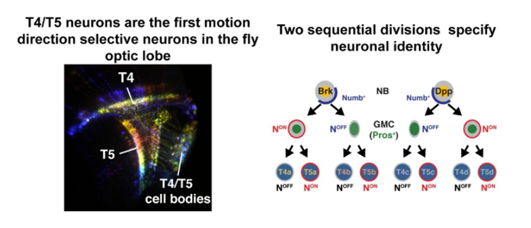
T4 and T5 have four subtypes, each responding to motion in one of four directions (back to front, front to back as well as up and down), such that all T4 and T5 neurons with the same directional preference work to support the perception of self-motion.
In their study, the scientists discovered that T4 and T5 neurons originate from two pools of stem cells—one that produces cells sensitive to horizontal motion and the other attuned to vertical motion.
These stem cells, they found, then rely on a novel mode of neuron production where two sequential cell divisions produce sets of two T4 and two T5 neurons with opposite motion direction selectivity—for instance, one T4/T5 pair is sensitive to back-to-front movement and the other to front-to-back.
“Because T4 and T5 neurons with opposite motion direction selectivity are produced by the same stem cell at the same time, these four neurons synchronously project to the same position in the brain corresponding to a specific point in visual space,” explains Desplan. “As a result, the organization of neuronal projections results directly from neuronal birth order, illustrating how simple developmental rules can produce complex neural organization.”
Funding: The study was supported by a grant from the CGSB of the NYU Abu Dhabi Institute and by the National Institutes of Health (R01 EY017916).
Source: James Devitt – NYU
Publisher: Organized by NeuroscienceNews.com.
Image Source: NeuroscienceNews.com image is credited to Filipe Pinto-Teixeira.
Video Source: Video credited to Filipe Pinto Teixeira Sousa.
Original Research: Abstract in Cell.
doi:10.1016/j.cell.2018.02.053
[cbtabs][cbtab title=”MLA”]NYU “Shedding Light on the Perception of Motion.” NeuroscienceNews. NeuroscienceNews, 22 March 2018.
<https://neurosciencenews.com/motion-perception-8676/>.[/cbtab][cbtab title=”APA”]NYU (2018, March 22). Shedding Light on the Perception of Motion. NeuroscienceNews. Retrieved March 22, 2018 from https://neurosciencenews.com/motion-perception-8676/[/cbtab][cbtab title=”Chicago”]NYU “Shedding Light on the Perception of Motion.” https://neurosciencenews.com/motion-perception-8676/ (accessed March 22, 2018).[/cbtab][/cbtabs]
Abstract
Development of Concurrent Retinotopic Maps in the Fly Motion Detection Circuit
Highlights
•A neuroblast produces four neurons detecting two directions of bright or dark edge motion
•Distinct progenitors produce neurons processing vertical versus horizontal motion
•Notch specifies neurons responding to bright or dark edge motion
•This mode of neurogenesis establishes retinotopy in the fly global motion system
Summary
Understanding how complex brain wiring is produced during development is a daunting challenge. In Drosophila, information from 800 retinal ommatidia is processed in distinct brain neuropiles, each subdivided into 800 matching retinotopic columns. The lobula plate comprises four T4 and four T5 neuronal subtypes. T4 neurons respond to bright edge motion, whereas T5 neurons respond to dark edge motion. Each is tuned to motion in one of the four cardinal directions, effectively establishing eight concurrent retinotopic maps to support wide-field motion. We discovered a mode of neurogenesis where two sequential Notch-dependent divisions of either a horizontal or a vertical progenitor produce matching sets of two T4 and two T5 neurons retinotopically coincident with pairwise opposite direction selectivity. We show that retinotopy is an emergent characteristic of this neurogenic program and derives directly from neuronal birth order. Our work illustrates how simple developmental rules can implement complex neural organization.


