

Summary: A new electronic microscopy study has captured microglia performing their role in synaptic pruning.
Source: EMBL.
For the first time, EMBL researchers have captured microglia nibbling on brain synapses. Their findings show that the special glial cells help synapses grow and rearrange, demonstrating the essential role of microglia in brain development. Nature Communications will publish the results on March 26.
Around one in ten cells in your brain are microglia. Cousins of macrophages, they act as the first and main contact in the central nervous system’s active immune defense. They also guide healthy brain development. Researchers have proposed that microglia pluck off and eat synapses – connections between brain cells – as an essential step in the pruning of connections during early circuit refinement. But, until now, no one had seen them do it.
Microglia make synapses stronger
That is why Laetitia Weinhard, from the Gross group at EMBL Rome, set out on a massive imaging study to actually see this process in action in the mouse brain, in collaboration with the Schwab team at EMBL Heidelberg. “Our findings suggest that microglia are nibbling synapses as a way to make them stronger, rather than weaker,” says Cornelius Gross, who led the work.
Warm welcome
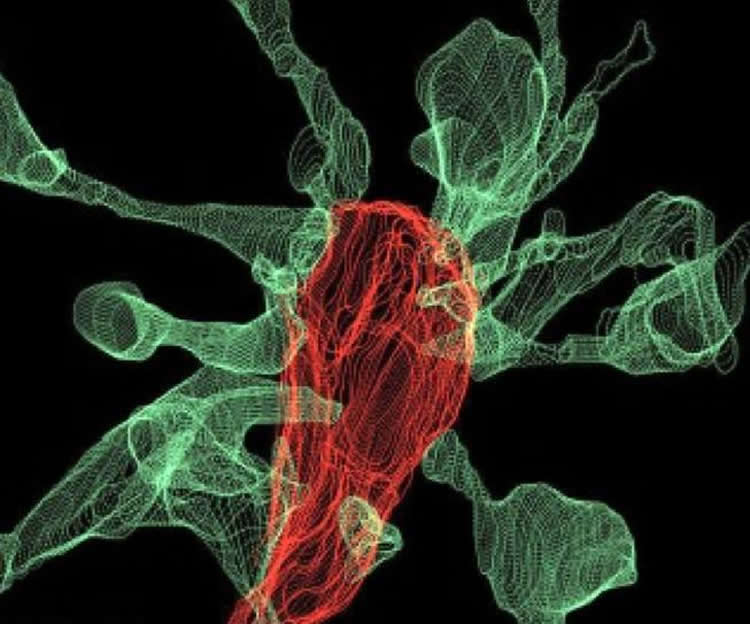
The team saw that around half of the time that microglia contact a synapse, the spine head sends out thin projections or ‘filopodia’ to greet them. In one particularly dramatic case – as seen in the accompanying image – fifteen spine heads extended filopodia toward a single microglia as it picked on a synapse. “As we were trying to see how microglia eliminate synapses, we realised that microglia actually induce their growth most of the time,” Laetitia Weinhard explains.
It turns out that microglia might underly the formation of double synapses, where the terminal end of a neuron releases neurotransmitters onto two neighboring partners instead of one. This process can support effective connectivity between neurons. Weinhard: “This shows that microglia are broadly involved in structural plasticity and might induce the rearrangement of synapses, a mechanism underlying learning and memory.”
Perseverance
Since this was the first attempt to visualise this process in the brain, the current paper entails five years of technological development. The team tried three different state-of-the-art imaging systems before she succeeded. Finally, by combining correlative light and electron microscopy (CLEM) and light sheet fluorescence microscopy – a technique developed at EMBL – they were able to make the first movie of microglia eating synapses.
“This is what neuroscientists fantasised about for years, but nobody had ever seen before,” says Cornelius Gross. “These findings allow us to propose a mechanism for the role of microglia in the remodeling and evolution of brain circuits during development.” In the future, he plans to investigate the role of microglia in brain development during adolescence and the possible link to the onset of schizophrenia and depression.
Source: Iris Kruijen – EMBL
Publisher: Organized by NeuroscienceNews.com.
Image Source: NeuroscienceNews.com image is credited to L. Weinhard, EMBL Rome.
Original Research: Open access research for “Microglia remodel synapses by presynaptic trogocytosis and spine head filopodia induction” by Laetitia Weinhard, Giulia di Bartolomei, Giulia Bolasco, Pedro Machado, Nicole L. Schieber, Urte Neniskyte, Melanie Exiga, Auguste Vadisiute, Angelo Raggioli, Andreas Schertel, Yannick Schwab & Cornelius T. Gross in Nature Communications. Published online March 26 2018.
doi:10.1038/s41467-018-03566-5
[cbtabs][cbtab title=”MLA”]EMBL “Captured on Film For the First Time: Microglia Nibbling on Synapses.” NeuroscienceNews. NeuroscienceNews, 26 March 2018.
<https://neurosciencenews.com/microglia-synapses-8685/>.[/cbtab][cbtab title=”APA”]EMBL (2018, March 26). Captured on Film For the First Time: Microglia Nibbling on Synapses. NeuroscienceNews. Retrieved March 26, 2018 from https://neurosciencenews.com/microglia-synapses-8685/[/cbtab][cbtab title=”Chicago”]EMBL “Captured on Film For the First Time: Microglia Nibbling on Synapses.” https://neurosciencenews.com/microglia-synapses-8685/ (accessed March 26, 2018).[/cbtab][/cbtabs]
Microglia remodel synapses by presynaptic trogocytosis and spine head filopodia induction
Microglia are highly motile glial cells that are proposed to mediate synaptic pruning during neuronal circuit formation. Disruption of signaling between microglia and neurons leads to an excess of immature synaptic connections, thought to be the result of impaired phagocytosis of synapses by microglia. However, until now the direct phagocytosis of synapses by microglia has not been reported and fundamental questions remain about the precise synaptic structures and phagocytic mechanisms involved. Here we used light sheet fluorescence microscopy to follow microglia–synapse interactions in developing organotypic hippocampal cultures, complemented by a 3D ultrastructural characterization using correlative light and electron microscopy (CLEM). Our findings define a set of dynamic microglia–synapse interactions, including the selective partial phagocytosis, or trogocytosis (trogo-: nibble), of presynaptic structures and the induction of postsynaptic spine head filopodia by microglia. These findings allow us to propose a mechanism for the facilitatory role of microglia in synaptic circuit remodeling and maturation.


