
Summary:
Researchers at the University of Oxford have discovered that human microglia can selectively prune away toxic alpha-synuclein aggregates from dopamine-producing neurons without destroying the host nerve cells. Utilizing co-cultures of human induced pluripotent stem cell (iPSC)-derived neurons and microglia, the team demonstrated that microglia employ “trogocytosis”, a precise cellular nibbling mechanism, regulated by GPNMB, P2RY12, and CD22 signaling pathways, along with an IL-10 molecular brake. The findings identify a distinct, neuroprotective microglial subtype that shields neurons in Parkinson’s disease.
Key Facts:
- Selective Pruning via Trogocytosis: Human microglia selectively extract and dispose of aggregated alpha-synuclein clumps by “nibbling” specific segments of living dopamine neurons while preserving overall neuronal viability and connectivity.
- GPNMB Identified as Functional Driver: Glycoprotein non-metastatic melanoma protein B (GPNMB), a protein previously linked to Parkinson’s risk via genome-wide association studies (GWAS), binds directly to alpha-synuclein and is required for microglia to effectively clear aggregates.
- Fine-Tuned Immune Brakes: The protective clearance mechanism is controlled by sensing receptors (P2RY12), inhibitory checkpoints (CD22), and an interleukin-10 (IL-10) autocrine brake that prevents runaway neurotoxic inflammation.
Source: University of Oxford
Beyond the Double-Edged Sword: A Precision Immune Defense
In neurodegenerative diseases, microglia, the central nervous system’s resident immune sentinels, are typically framed through a conflicted narrative. Under physiological conditions, they survey brain tissue, prune synapses, and clear cellular debris; during chronic neurodegeneration, their persistent activation drives inflammatory signaling cascades that accelerate neuronal death.
Nowhere has this paradox been more evident than in Parkinson’s disease, a condition affecting more than 10 million individuals globally. The disease is defined by the selective death of dopamine-producing neurons within the substantia nigra, driven largely by the misfolding and aggregation of alpha-synuclein into toxic intracellular clumps.
Whether human microglia can intervene constructively to rescue dopamine neurons from this proteinaceous burden, without destroying the neurons in the process, has remained an open question.
Now, a team of neuroscientists at the University of Oxford has revealed an unexpected, highly targeted defense mechanism.
The researchers established that a specialized population of human microglia actively extracts and destroys harmful alpha-synuclein aggregates directly from living dopamine neurons via trogocytosis (from the Greek trogo, meaning “to gnaw” or “to nibble”).
“What is striking is the precision of this response,” said first author Hung-Ju Chueh, Ph.D., of the University of Oxford. “The microglia were not simply engulfing damaged neurons but instead removing parts of the neuron containing aggregated alpha-synuclein, suggesting that, at certain stages of disease, microglia help neurons dispose of potentially harmful material.”
Engineering Human iPSC Co-Cultures to Model Live Interactions
To investigate interactions between human brain cells in real time, the Oxford investigators generated human induced pluripotent stem cell (iPSC) lines, co-culturing authentic human dopamine neurons alongside human microglia.
The team modeled alpha-synuclein pathology through two complementary methods: by introducing genetic alpha-synuclein gene dosage elevations (triplication), and by seeding the cultures with exogenous preformed alpha-synuclein fibrils that template the misfolding of native neuronal protein.
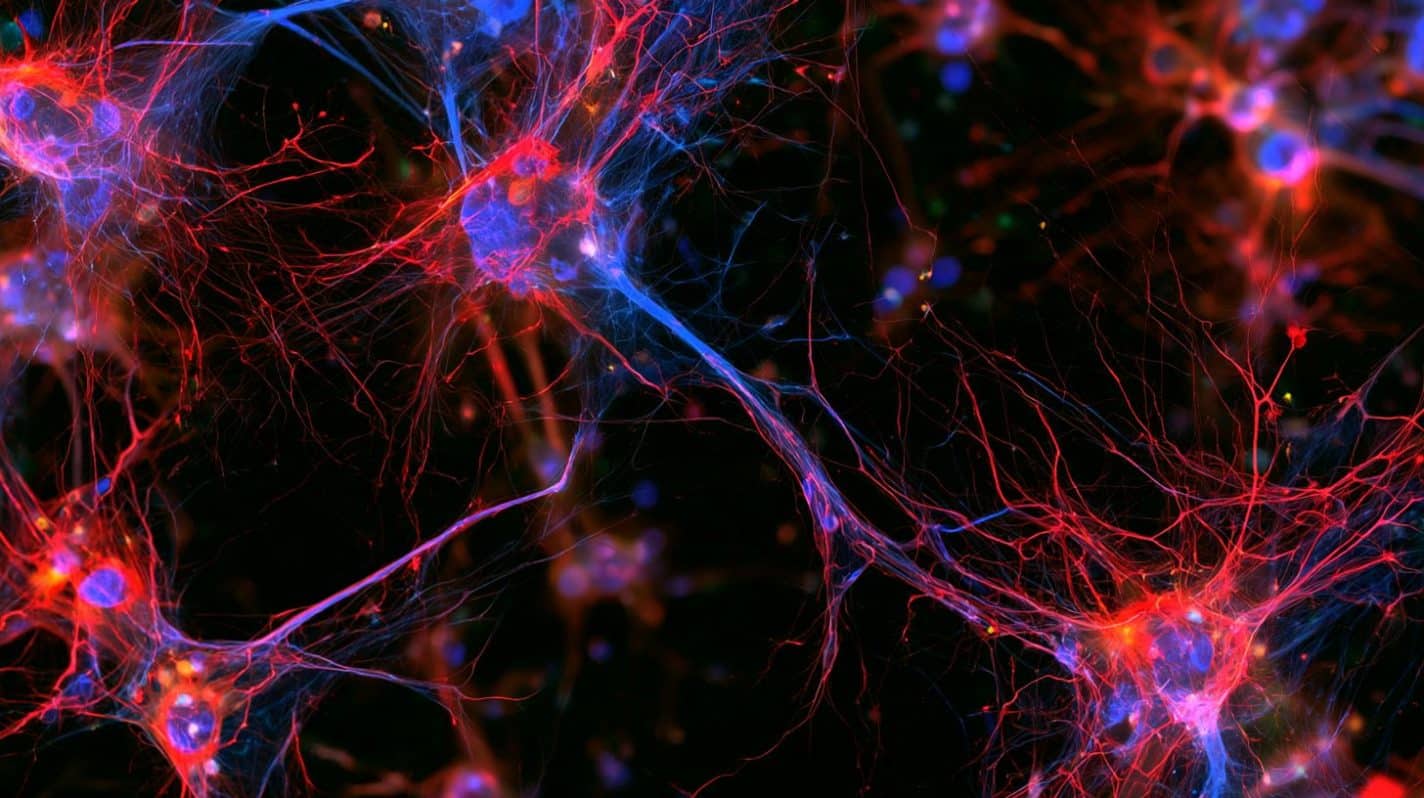
High-resolution cellular imaging revealed that rather than engulfing entire dying neurons via standard phagocytosis, the microglia selectively nibbled off membrane pockets enclosing the aggregated protein. This process of trogocytosis eliminated the toxic pathology while leaving the broader axonal network and cell body intact.
Single-cell RNA sequencing showed that this clearance was driven by a distinct, transcriptionally defined subpopulation of activated microglia. This subset was kept from over-activating through a balanced signaling network:
- Surveillance and Guidance: Governed by P2RY12 purinergic receptor sensing.
- Cellular Checkpoints: Regulated by CD22 inhibitory surface interactions between neurons and microglia.
- Inflammatory Suppression: Maintained by an autocrine interleukin-10 (IL-10) brake that halted secondary, collateral inflammatory tissue injury.
Solving the GPNMB Genetic Mystery in Parkinson’s
The investigation also cracked a longstanding genetic puzzle. Multiple large-scale genome-wide association studies (GWAS) had previously flagged variants in the GPNMB (glycoprotein non-metastatic melanoma protein B) gene locus as significant risk factors for Parkinson’s disease, but its mechanistic function in the brain remained unknown.
The Oxford team discovered that GPNMB is directly upregulated in human microglia when exposed to aggregate-bearing neurons, where it physically interacts with pathological alpha-synuclein within the microglial machinery.
Furthermore, post-mortem analysis of human brain tissue from patients with incidental Lewy body disease and confirmed Parkinson’s disease revealed elevated GPNMB levels within substantia nigra microglia.
To confirm causality, the team used CRISPR interference (CRISPRi) to knock down GPNMB expression in human microglia. Deprived of GPNMB, the microglia lost their ability to clear alpha-synuclein aggregates from the neighboring dopamine neurons, establishing GPNMB as an essential driver of the neuroprotective response.
“Our findings highlight that the immune response in Parkinson’s disease is more nuanced than simply being beneficial or harmful,” noted senior author George Tofaris, M.D., Ph.D., Professor of Neurology and Translational Neuroscience at the University of Oxford. “We have identified a population of human microglia that can actively remove pathological alpha-synuclein from neurons. Understanding how to enhance and monitor such beneficial microglial functions, without triggering damaging inflammation, could open up new avenues for developing disease-modifying treatments.”
Editorial Notes:
- This article was edited by a Neuroscience News editor.
- Journal paper reviewed in full.
- Additional context added by our staff.
About this Neurology Research:
- Media Contact: Christopher McIntyre
- Source: University of Oxford
- Image Credit: Image credited to Neuroscience News
- Original Research is Open Access: Science Translational Medicine (Oct 7, 2026). “Human microglia clear intraneuronal alpha- synuclein aggregates by GPNMB-mediated trogocytosis.” Authors: Hung-Ju Chueh, Antigoni Katsikoudi, Ana Aragón-González, Chor Lai Lam, Liezel Tamon, Ashwin Jainarayanan, Devika Agarwal, Sally A. Cowley, David Sims, and George K. Tofaris.
- DOI: 10.1126/scitranslmed.adz9258
Abstract
Human microglia clear intraneuronal alpha- synuclein aggregates by GPNMB-mediated trogocytosis
Microglia are the primary immune cells of the brain, but their role in Parkinson’s disease is not fully understood. Chronic microglial activation is toxic to neurons, but in the early stages of pathology, microglia also exert beneficial functions.
We used induced pluripotent stem cell (iPSC)–derived coculture models to investigate how human microglia respond to α-synuclein aggregates that form de novo inside human dopaminergic neurons with α-synuclein gene triplication or are triggered by fibrils. We found that microglia cleared Ser129-phosphorylated α-synuclein aggregates through a contact-dependent mechanism.
This process was fine-tuned by sensing (P2RY12) and inhibitory (CD22) signals and involved selective phagocytosis of neuronal subcompartments (trogocytosis), independently of TREM2 or phosphatidylserine. In the presence of intraneuronal α-synuclein aggregates, microglia exhibited morphological and transcriptional changes indicative of activation.
Using single-cell sequencing, we identified a cluster of disease-associated microglia (DAM) responsible for this beneficial phenotype and the Parkinson’s disease GWAS candidate glycoprotein nonmetastatic melanoma protein B (GPNMB) as a key effector of aggregate clearance by microglial lysosomes. GPNMB interacted with Ser129-phosphorylated α-synuclein in iPSC-derived microglia exposed to aggregate-laden dopaminergic neurons and was up-regulated in substantia nigra microglia of individuals with incidental Lewy bodies or Parkinson’s disease.
Microglia-specific GPNMB knockdown using CRISPRi reduced intraneuronal aggregate clearance. GPNMB-mediated trogocytosis was negatively regulated by an autocrine IL-10 signaling loop, whereas IL-10 receptor blockade enhanced the phagocytic response. Together, our study identifies a subtype of human microglia capable of removing intraneuronal aggregates and potential therapeutic targets.