

Summary: Researchers have released a new open-access 3D map of the cellular structure of two distinct parts of the metathalamus.
Source: Human Brain Project
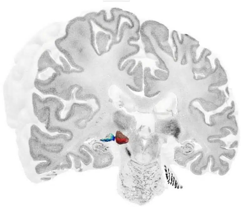
The human metathalamus is extremely important for relaying auditory and visual signals to the rest of the brain. However, a detailed, three-dimensional map of the cellular structure of the area was still missing.
Using the BigBrain dataset, an integral part of the HBP, the researchers focused on the two distinct parts of the metathalamus: the medial geniculate body (MGB) with its subdivisions; and lateral geniculate body (LGB) with its six layers.
“The BigBrain dataset helps us to understand the structure of complex subcortical nuclei,” explains Andrea Brandstetter of the Institute of Neuroscience and Medicine, Forschungszentrum Jülich, in a video by HBP partnering project HIBALL.
“Novel deep-learning-based approaches are used to learn about the topography and the cellular architecture of the metathalamus,” says Kai Kiwitz of the Cécile and Oskar Vogt Institute of Brain Research, University Hospital Düsseldorf.
The new 3D maps are freely available online as part of the EBRAINS Atlas.
“This way they can be used by the scientific community to bridge the microscale histology of BigBrain with functional measurements,” explains Kiwitz.
In addition to expanding our knowledge of the brain structure, the maps also have clinical relevance: many neurological disorders and dysfunctions involve the metathalamus, and highly detailed information regarding these structures could be used in conjunction with neuroimaging to better inform diagnosis and aid neurosurgery and deep brain stimulation.
About this brain mapping research news
Author: Press Office
Source: Human Brain Project
Contact: Press Office – Human Brain Project
Image: The image is credited to Kiwitz, Brandstetter et al, 2022
Original Research: Open access.
“Cytoarchitectonic Maps of the Human Metathalamus in 3D Space” by Kai Kiwitz et al. Frontiers in Neuroanatomy
Abstract
Cytoarchitectonic Maps of the Human Metathalamus in 3D Space
The human metathalamus plays an important role in processing visual and auditory information. Understanding its layers and subdivisions is important to gain insights in its function as a subcortical relay station and involvement in various pathologies. Yet, detailed histological references of the microanatomy in 3D space are still missing.
We therefore aim at providing cytoarchitectonic maps of the medial geniculate body (MGB) and its subdivisions in the BigBrain – a high-resolution 3D-reconstructed histological model of the human brain, as well as probabilistic cytoarchitectonic maps of the MGB and lateral geniculate body (LGB). Therefore, histological sections of ten postmortem brains were studied.
Three MGB subdivisions (MGBv, MGBd, MGBm) were identified on every 5th BigBrain section, and a deep-learning based tool was applied to map them on every remaining section. The maps were 3D-reconstructed to show the shape and extent of the MGB and its subdivisions with cellular precision. The LGB and MGB were additionally identified in nine other postmortem brains.
Probabilistic cytoarchitectonic maps in the MNI “Colin27” and MNI ICBM152 reference spaces were computed which reveal an overall low interindividual variability in topography and extent. The probabilistic maps were included into the Julich-Brain atlas, and are freely available. They can be linked to other 3D data of human brain organization and serve as an anatomical reference for diagnostic, prognostic and therapeutic neuroimaging studies of healthy brains and patients.
Furthermore, the high-resolution MGB BigBrain maps provide a basis for data integration, brain modeling and simulation to bridge the larger scale involvement of thalamocortical and local subcortical circuits.


