Summary: New research reveals that the insula, a key brain region, shows altered connectivity in people who struggle to understand speech in noisy environments. Using resting-state MRI, scientists found that the left insula works harder even when the brain is not actively processing sound, suggesting a permanent rewiring linked to hearing difficulties.
Since insula abnormalities are also associated with early dementia, these findings may help explain the established link between hearing loss and cognitive decline. Researchers also discovered that exposure to noisy environments may help train the brain, raising hope for improving hearing challenges through practice.
Key Facts:
- Insula Overactivation: People with speech-in-noise difficulties show stronger insula connectivity, even at rest.
- Link to Dementia: Insula rewiring may explain connections between hearing loss and cognitive decline.
- Potential for Improvement: Exposure to noisy environments may help train the brain to better process speech.
Source: University at Buffalo
As they age, some people find it harder to understand speech in noisy environments.
Now, University at Buffalo researchers have identified the area in the brain, called the insula, that shows significant changes in people who struggle with speech in noise.
The findings, published in the journal Brain and Language, contribute to the growing link between hearing loss and cognitive impairment leading to dementia.

Previous research has separately established connections between hearing difficulties and dementia, as well as insula abnormalities and cognitive decline.
The insulae are two complicated structures that interact with the brain’s frontal lobe, which is responsible for higher-level cognitive function. The insulae integrate sensory, emotional and cognitive information.
The study involved 40 men and women ages 20-80. They underwent hearing testing first to determine who had difficulty hearing speech in noisy environments; they then underwent magnetic resonance imaging (MRI) tests of their brains at rest.
The brain at baseline
While task-based studies reveal what parts of the brain activate during specific activities, the researchers in this study wanted to see how difficulties hearing speech in noise might affect the brain at baseline, at rest.
According to David S. Wack, PhD, first author and associate professor of radiology in the Jacobs School of Medicine and Biomedical Sciences at UB, resting-state MRI reveals functional connections, showing how different brain regions work together even when not actively engaged in tasks.
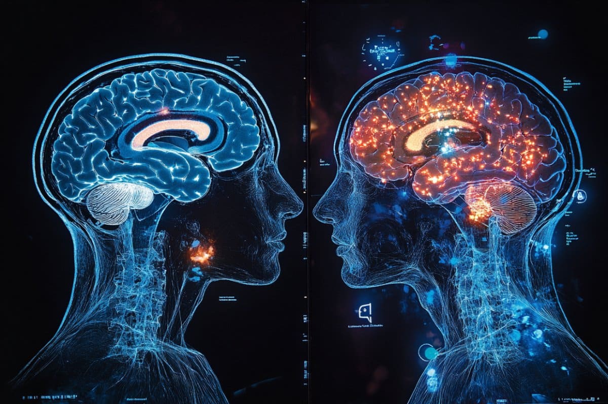
The study found that the left insula shows stronger connectivity with auditory regions in people who struggle with speech in noise, suggesting a permanent rewiring of brain networks that persists even when they’re not actively listening to challenging speech.
“Your brain is always doing something,” Wack explains.
“And when you have hearing loss, you are recruiting other areas of the brain to do more processing in order to decode what’s going on. What’s interesting is that we found the insula working harder when the brain was supposedly at rest, when there was no speech in noise.”
That finding, he says, has implications for how dementia may develop, since the insula is also associated with early dementia.
“Multiple studies have established correlations between hearing loss, speech-in-noise difficulties and dementia,” Wack explains.
“Our findings show baseline brain connectivity changes that occur with poorer performance with speech in noise that may explain these connections.
“You have a bad signal coming (the sound or the seen object) and then the brain is left to interpret what it means,” he says.
“Your brain fills in what works, what seems to make the most sense and it incorporates higher-level regions of the brain to do it.
He continues: “It’s not that hearing loss causes dementia, but if we could find a way to preserve the fidelity of the signal coming in, then the brain wouldn’t have to start compensating for that hearing loss.”
An unexpected finding
The researchers reported one unexpected and fascinating finding. “We had a subject who had relatively poor hearing for pure tones, but who achieved the highest score for speech-in-noise for one ear,” Wack says.
It turned out this person worked in an environment with high background noise.
“This suggests that people don’t have to just accept poor performance in noisy backgrounds,” says Wack.
“It shows that you might be able to practice your way out of it.”
He hopes to further study the relationship between hearing loss and dementia. “By identifying shared neural networks at rest, our research adds to the understanding of why addressing hearing difficulties might help with cognitive function,” he says.
University at Buffalo co-authors include Ferdinand Schweser, PhD, of the Department of Neurology; Sarah F. Muldoon, PhD, of the Department of Mathematics in the College of Arts and Sciences; and Robert S. Miletich, MD, PhD, formerly interim chair and professor of nuclear medicine.
Kathleen McNerney, PhD, from SUNY Buffalo State, also contributed to this work. Other co-authors are from the Boston University School of Medicine, the Institute of Science in Tokyo and Canon Medical Systems.
The imaging work was done at the Center for Biomedical Imaging in UB’s Clinical and Translational Science Institute.
Funding: Funding for the study and resources were provided by Canon Medical Systems USA, the National Center for Advancing Translational Sciences (NIH, UL1TR001412) and by a gift from William and Grace Mabie for the advancement of neuroscience.
About this auditory neuroscience research news
Author: Ellen Goldbaum
Source: University at Buffalo
Contact: Ellen Goldbaum – University at Buffalo
Image: The image is credited to Neuroscience News
Original Research: Open access.
“Speech in noise listening correlates identified in resting state and DTI MRI images” by David S. Wack et al. Brain and Language
Abstract
Speech in noise listening correlates identified in resting state and DTI MRI images
This study presents an examination of the neural connectivity associated with processing speech in noisy environments, an ability that declines with age.
We correlated subjects’ speech-in-noise (SIN) ability with resting-state MRI scans and Fractional Anisotropy (FA) values from the auditory section of the corpus callosum, both with and without correcting for age.
The results revealed that subjects who performed poorly on the right ear SIN test (QuickSIN, MedRx) had higher correlations between the primary auditory cortex and regions of the brain that process language.
Subjects who performed well on the QuickSIN test had stronger correlations bilaterally between the primary auditory cortices, however, this finding was due to age. Likewise, FA values seem best explained by age not SIN.
The Ig2 region of the insula showed significant correlation with right ear SIN when correcting for age.


