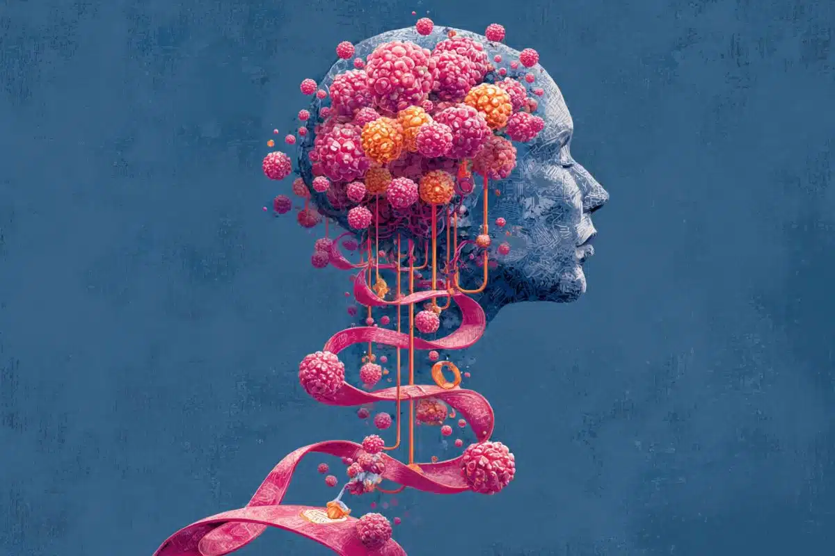
Young people with behavioural problems, such as antisocial and aggressive behaviour, show reduced grey matter volume in a number of areas of the brain, according to a new study published in JAMA Psychiatry.
The researchers from the University of Birmingham found that, compared to typically developing youths, those with behavioural problems show grey matter reductions specifically within the amygdala, the insula, and the prefrontal cortex.
These brain areas are important for decision-making, empathic responses, reading facial expressions and emotion regulation; key cognitive and affective processes that are shown to be deficient in youths with behavioural problems.
The article combined brain imaging data from 13 existing studies including 394 youths with behavioural problems and 350 typically developing youths, making it the largest study on this topic.
Dr Stephane De Brito, lead author of the paper, explained, “We know that severe behavioural problems in youths are not only predictive of antisocial and aggressive behaviour in adulthood, but also substance misuse, mental health problems and poor physical health.”
“For that reason, behavioural problems are an essential target for prevention efforts and our study advances understanding of the brain regions associated with aggressive and antisocial behaviour in youths.”
However, a number of unanswered questions still remain in the field. For example, the extent to which these structural differences in the brain are associated with environmental factors (such as smoking/substance abuse during pregnancy and maltreatment in early childhood) is still poorly understood.
Dr Jack Rogers, Research Fellow at the University of Birmingham, said, “There are a lot of questions still outstanding. For instance, prospective longitudinal studies are needed to assess if these structural differences are present early in life and if they persist over a longer period of time.”

“In future research, it will also be important to examine if these brain differences, and the affective and cognitive processes they are involved in, can be influenced by therapeutic interventions to promote a good outcome in adult life.”
Dr De Brito noted, “Some of those important questions will be addressed in the context of a large multisite study we are involved in. This research will be carried out on children and adolescents from seven European countries to examine the environmental and neurobiological factors implicated in the development of behavioural problems in male and female youths.”
Source: Luke Harrison – University of Birmingham
Image Source: The image is adapted from the University of Birmingham press release
Original Research: Full open access research for “Cortical and Subcortical Gray Matter Volume in YouthsWith Conduct Problems” by Jack C. Rogers, PhD and Stéphane A. De Brito, PhD in JAMA Psychiatry. Published online December 9 2015 doi:10.1001/jamapsychiatry.2015.2423
Abstract
Cortical and Subcortical Gray Matter Volume in YouthsWith Conduct Problems
Importance A large number of structural neuroimaging studies have used voxel-based morphometry (VBM) to identify gray matter abnormalities in youths with conduct problems (CP), but the findings have been disparate and few have been replicated.
Objective To conduct a meta-analysis of published whole-brain structural neuroimaging studies on youths with CP that used VBM methods to facilitate replication and aid further analyses by researchers.
Data Sources The PubMed, ScienceDirect, Scopus, Google Scholar, and Web of Science databases were searched for VBM studies published from January 1, 2007, through March 31, 2015. Manual searches were conducted using title and citation information. Authors were contacted for additional data.
Study Selection A literature search identified 28 studies. Studies were excluded if they (1) failed to use VBM, (2) failed to report a voxelwise comparison between youths with CP and typically developing (TD) youths, (3) used different significance or extent thresholds throughout the brain, (4) included duplicated datasets, and (5) did not provide peak coordinates or parametric maps after contact with the authors. Thirteen studies were deemed eligible for inclusion (394 youths with CP and 350 TD youths).
Data Extraction and Synthesis Anisotropic effect-size signed differential mapping (SDM) was used for voxel-based meta-analyses. Statistical parametric maps comparing gray matter differences between youths with CP and TD youths were available for 11 of the studies, with peak coordinates available for the remaining studies.
Main Outcomes and Measures Regional gray matter volume (GMV) differences in youths with CP compared with TD youths.
Results Youths with CP had decreased GMV in the left amygdala (SDM estimate = −0.218; P < .001) (extending into anterior insula), right insula (SDM estimate = −0.174; P < .001) (extending ventrolaterally into the prefrontal cortex and inferiorly into the superior temporal gyrus), left medial superior frontal gyrus (SDM estimate = −0.163; P = .001) (extending into the right anterior cingulate cortex), and left fusiform gyrus (SDM estimate = −0.146; P = .003). Subgroup meta-analysis assessing age-at-onset effects identified reduced GMV in the left amygdala (SDM estimate = −0.232; P < .001) (extending into anterior insula). Meta-regression analyses revealed that greater scores on measures of callous-unemotional traits were associated with a lower reduction in GMV in the left putamen (SDM estimate = −0.911; P < .001). The proportion of male and female youths in the sample was associated with decreased GMV in the left amygdala (SDM estimate = −0.31; P < .001) and increased GMV in the right inferior temporal cortex (SDM estimate = 0.755; P < .001). While there was no association with comorbid attention-deficit/hyperactivity disorder or IQ, age range was associated with gray matter differences in the left amygdala.
Conclusions and Relevance We identified gray matter reductions within the insula, amygdala, frontal and temporal regions in youths with CP as well as inconsistencies in sample characteristics across studies that should be addressed in future research.
“Cortical and Subcortical Gray Matter Volume in YouthsWith Conduct Problems” by Jack C. Rogers, PhD and Stéphane A. De Brito, PhD in JAMA Psychiatry. Published online December 9 2015 doi:10.1001/jamapsychiatry.2015.2423