

Summary: A new study looks at the role glutamate plays in neuromuscular development.
Source: University at Buffalo.
For decades, scientists thought acetylcholine was the only neurotransmitter responsible for controlling how muscles and nerves are wired together during development.
Turns out, they were wrong. Glutamate, the most common neurotransmitter in the brain, is also necessary.
Researchers at the University at Buffalo and Johns Hopkins University reported their findings with mice in the Journal of Neuroscience.
The team took a new approach to the old question of how the connections from the spinal cord to the muscles mature, says Kirkwood Personius, the paper’s lead author and a clinical associate professor of rehabilitation science in UB’s School of Public Health and Health Professions.
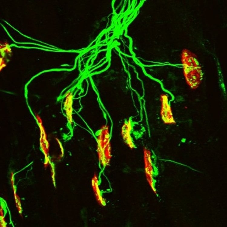
Each muscle is made of many individual muscle fibers and, in adults, each of those muscle fibers is contacted by a single motor neuron. However, this simple arrangement is not what you see at birth. Instead, each muscle fiber is contacted by as many as 10 nerves.
The process that allows one motor neuron to stay while all the others are retracted seems to be this, according to the researchers: the nerve that’s most effective in activating the muscle is the one that wins.
But what specifically occurs during the nerve’s firing that triggers the stabilizing of the winner and the withdrawal of the others? For many decades, it was assumed that the pruning process began with release of the neurotransmitter acetylcholine from the nerve. This seemed logical, the paper’s authors say, because motor neurons do indeed release lots of acetylcholine.
“However, we now have shown that an important transmitter is one that nobody had previously expected: it is glutamate,” said Personius, PT, PhD. “The nerves release a molecule that is converted into glutamate, and the glutamate then activates glutamate receptors, notably NMDA receptors, on the muscle.”
NMDA (N-methyl-D-aspartate) receptors are one of several types of molecules that respond to glutamate. They are especially important in the central nervous system controlling brain development, learning and synaptic plasticity. “Nobody thought NMDA receptors played any role in the innervation of muscle,” Personius said.
The researchers tested their hypothesis — that glutamate receptor activation modulates the development of the neuromuscular system — in several ways, each of which supported what they thought. In addition, they showed that the response of the muscle to glutamate is very strong at birth, but quickly disappears as mice mature.
“Our work restarts a field that was stuck because of the widespread conviction that the process depended on a single transmitter, acetylcholine,” said Susan Udin, PhD, a paper co-author and professor of physiology in UB’s Jacobs School of Medicine and Biomedical Sciences.
“This study opens up a wide range of experimental possibilities because so much is known from central nervous system studies about how NMDA receptors work. Our work opens up a possible understanding of why return of muscle function is often limited after peripheral nerve trauma.”
The same processes that control muscle fiber development tend to recur after peripheral injury in adults. Now, the research team is testing the hypothesis that the poor outcomes often seen after peripheral nerve trauma could be improved by manipulating NMDA receptors.
Barbara S. Slusher, PhD, professor of neurology, medicine, psychiatry and neuroscience at Johns Hopkins University, is also a paper co-author.
Source: David J. Hill – University at Buffalo
Image Source: NeuroscienceNews.com image is adapted from the University at Buffalo press release.
Original Research: Abstract for “Neuromuscular NMDA Receptors Modulate Developmental Synapse Elimination” by Kirkwood E. Personius1,2, Barbara S. Slusher4, and Susan B. Udin in Journal of Neuroscience. Published online August 24 2016 doi:10.1523/JNEUROSCI.1181-16.2016
[cbtabs][cbtab title=”MLA”]University at Buffalo. “Glutamate Plays Previously Unknown Role in Neuromuscular Development.” NeuroscienceNews. NeuroscienceNews, 19 September 2016.
<https://neurosciencenews.com/glutamate-neuromuscular-development-5074/>.[/cbtab][cbtab title=”APA”]University at Buffalo. (2016, September 19). Glutamate Plays Previously Unknown Role in Neuromuscular Development. NeuroscienceNews. Retrieved September 19, 2016 from https://neurosciencenews.com/glutamate-neuromuscular-development-5074/[/cbtab][cbtab title=”Chicago”]University at Buffalo. “Glutamate Plays Previously Unknown Role in Neuromuscular Development.” https://neurosciencenews.com/glutamate-neuromuscular-development-5074/ (accessed September 19, 2016).[/cbtab][/cbtabs]
Abstract
Neuromuscular NMDA Receptors Modulate Developmental Synapse Elimination
At birth, each mammalian skeletal muscle fiber is innervated by multiple motor neurons, but in a few weeks, all but one of those axons retracts (Redfern, 1970) and differential activity between inputs controls this phenomenon (Personius and Balice-Gordon, 2001; Sanes and Lichtman, 2001; Personius et al., 2007; Favero et al., 2012). Acetylcholine, the primary neuromuscular transmitter, has long been presumed to mediate this activity-dependent process (O’Brien et al., 1978), but glutamatergic transmission also occurs at the neuromuscular junction (Berger et al., 1995; Grozdanovic and Gossrau, 1998; Mays et al., 2009). To test the role of neuromuscular NMDA receptors, we assessed their contribution to muscle calcium fluxes in mice and tested whether they influence removal of excess innervation at the end plate. Developmental synapse pruning was slowed by reduction of NMDA receptor activation or expression and by reduction of glutamate production. Conversely, pruning is accelerated by application of exogenous NMDA. We also found that NMDA induced increased muscle calcium only during the first 2 postnatal weeks. Therefore, neuromuscular NMDA receptors play previously unsuspected roles in neuromuscular activity and synaptic pruning during development.
SIGNIFICANCE STATEMENT In normal adult muscle, each muscle fiber is innervated by a single axon, but at birth, fibers are multiply innervated. Elimination of excess connections requires neural activity; because the neuromuscular junction (NMJ) is a cholinergic synapse, acetylcholine has been assumed to be the critical mediator of activity. However, glutamate receptors are also expressed at the NMJ. We found that axon removal in mice is slowed by pharmacological and molecular manipulations that decrease signaling through neuromuscular NMDA receptors, whereas application of exogenous NMDA at the NMJ accelerates synapse elimination and increases muscle calcium levels during the first 2 postnatal weeks. Therefore, neuromuscular NMDA receptors play previously unsuspected roles in neuromuscular activity and elimination of excess synaptic input during development.
“Neuromuscular NMDA Receptors Modulate Developmental Synapse Elimination” by Kirkwood E. Personius1,2, Barbara S. Slusher4, and Susan B. Udin in Journal of Neuroscience. Published online August 24 2016 doi:10.1523/JNEUROSCI.1181-16.2016


