

Case Western Reserve University School of Medicine finding holds promise for repairing damaged hearing.
Hearing is made possible when hair bundles protruding from the tops of hair cells capture the energy of sound waves, converting them into electrical signals that stimulate the auditory nerve to the brain. These hair bundles are made up of individual hair-like projections, or stereocilia, which sway in unison with other stereocilia, and remain permanently with us throughout our lives. Extremely loud noise can force whipsaw motion of the stereocilia, causing them to be damaged and the resulting hearing loss to be permanent. The prevailing theory had been that these hair bundles were made up of cellular scaffolding proteins that do not change or circulate.
Scientists at Case Western Reserve University School of Medicine have discovered that the movement of protein within hair cells of the inner ear shows signs of a repair and renewal mechanism.
The investigator’s findings will be showcased as the cover paper in the Nov. 17 edition of Cell Reports and are available now in the online edition of the journal. The discovery paves the way for one day therapeutically manipulating the movement of these proteins in a way that could reverse hearing loss, a condition that affects approximately 15 percent of Americans between the ages of 20 and 69.
“What was surprising in our research with zebrafish is that proteins move so rapidly, implying that protein movement may be required to maintain the integrity of hair bundles in the inner ear,” said senior author Brian McDermott, PhD, associate professor of Otolaryngology-Head and Neck Surgery, Case Western Reserve University School of Medicine. “Our research tells us that constant movement, replacement and adjustment among proteins in the inner ear’s hair bundles serve a maintenance and even repair function.”
In their research, School of Medicine investigators discovered that proteins in the hair bundle move at surprisingly fast rates, and this movement could explain why hair bundles last a lifetime. The researchers used a confocal microscope to examine the movement in real time of these proteins within the inner ears of baby zebrafish. The zebrafish at this early state of development are completely transparent, making it possible to view internal organs without dissection, including ear structure.
“We made movies of the secret inner workings of the hair bundle in a live animal, and what is happening in the ear is amazing and unexpected,” McDermott said.
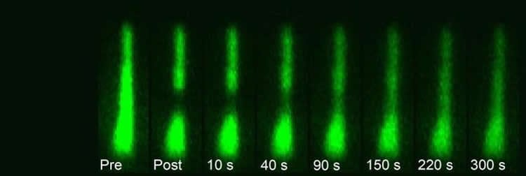
The researchers discovered that vital proteins, actin and myosin, move at a leisurely pace over the course of hours. On the other hand, fascin 2b moves incredibly fast, in seconds. This research proved that the internal structure of stereocilia is dynamic, that the proteins move frequently rather than being mostly stationary.
The speed with which fascin 2b moves may facilitate repair of breaks in the actin cables of the hair bundle when the ear is exposed to loud noise. Sound enters the ear at remarkable speeds, causing intense vibrations and back-and-forth movement of the hair bundle at extremely high rates. Investigators found that what appears to make the hair bundle so durable in the face of such forces is the rate of protein turnover, which enables renewal of the hair bundle.
“It was once thought that most everything within the stereocilia was relatively immobile and static,” McDermott said. “We found that the constant, dynamic movement likely contributes to the permanency of the hair bundle structure to last a lifetime, or 70 to 90 years in human terms.”
As a next research step, McDermott’s team will determine how the hair bundle may repair itself with such protein movement. The process likely involves fascin 2b but may also involve other proteins. They will continue to visualize this process fully in the transparent zebrafish ear.
“No one has shown that stereocilia heal themselves, so next we will find out if there is a repair mechanism,” he said. “People go deaf from damage to stereocilia in the inner ear. If we figure out ways of manipulating protein dynamics within stereocilia to heal this damage, we would likely be able to diminish hearing loss in humans.”
Joining McDermott in this research were lead coauthors Philsang Hwang and Shih-Wei Chou and contributing author Zongwei Chen.
Funding: This research was funded by National Institutes of Health grants DC009437 and S10RR017980 and by the Center for Clinical Research and Technology at University Hospitals Case Medical Center.
Source: Jeannette Spalding – Case Western Reserve
Image Credit: The image is credited to Cell Reports: Adapted from Figure 1 in “The Stereociliary Paracrystal is a Dynamic Cytoskeletal Scaffold In Vivo”
Original Research: Full open access research for “The Stereociliary Paracrystal Is a Dynamic Cytoskeletal Scaffold In Vivo” by Philsang Hwang, Shih-Wei Chou, Zongwei Chen, and Brian M. McDermott Jr. in Cell Reports. Published online November 5 2015 doi:10.1016/j.celrep.2015.10.003
Abstract
The Stereociliary Paracrystal Is a Dynamic Cytoskeletal Scaffold In Vivo
Highlights
•Transgenic zebrafish reveal stereociliary protein dynamics in vivo
•Actin cross-linker fascin 2b exchange within the stereocilium is rapid and unbiased
•Stereocilia exchange β-actin and myosin XVa in the shafts and tips, respectively
•Protein dynamism is suggested to underpin stereociliary function and longevity
Summary
Permanency of mechanosensory stereocilia may be the consequence of low protein turnover or rapid protein renewal. Here, we devise a system, using optical techniques in live zebrafish, to distinguish between these mechanisms. We demonstrate that the stereocilium’s abundant actin cross-linker fascin 2b exchanges, without bias or a phosphointermediate, orders of magnitude faster (t1/2 of 76.3 s) than any other known hair bundle protein. To establish the logic of fascin 2b’s exchange, we examine whether filamentous actin is dynamic and detect substantial β-actin exchange within the stereocilium’s paracrystal (t1/2 of 4.08 hr). We propose that fascin 2b’s behavior may enable cross-linking at fast timescales of stereocilia vibration while noninstructively facilitating the slower process of actin exchange. Furthermore, tip protein myosin XVa fully exchanges in hours (t1/2 of 11.6 hr), indicating that delivery of myosin-associated cargo occurs in mature stereocilia. These findings suggest that stereocilia permanency is underpinned by vibrant protein exchange.
“The Stereociliary Paracrystal Is a Dynamic Cytoskeletal Scaffold In Vivo” by Philsang Hwang, Shih-Wei Chou, Zongwei Chen, and Brian M. McDermott Jr. in Cell Reports. Published online November 5 2015 doi:10.1016/j.celrep.2015.10.003


