

Summary: Researchers report visual features modulated by the optokinetic reflex are encoded in the retina.
Source: LMU.
Contrast has an impact on the optokinetic reflex, which enables us to clearly perceive the landscape from a moving train. LMU researchers have now shown that visual features that modulate this ability are encoded in the retina.
When we gaze out the window of a moving train, our eye muscles are constantly at work, stabilizing the gaze in order to keep the passing landscape in focus. This so-called optokinetic reflex allows us to gauge the relative velocity of the passing scene, and thus helps us to stabilize the rapidly changing images on the retina – otherwise we would perceive what moves past us beyond the carriage window as a diffuse blur. Researchers led by LMU neurobiologist Professor Hans Straka have now shown experimentally that visual image features of the environment play an important role in the analysis and assessment of relative motion. In particular, they find that the levels of contrast determine the efficiency with which the passing parade is perceived by a moving observer. The results of the new study appear in the Journal of Experimental Biology.
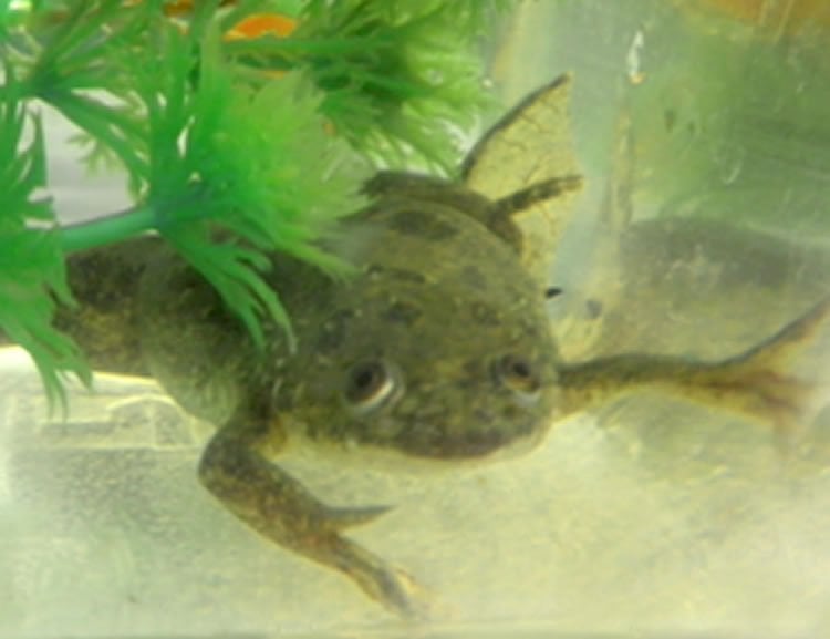
All vertebrates display optokinetic reflexes, which are responsible for fine-tuning the involuntary movements of the eye muscles, which in turn permit us to faithfully stabilize objects when we move. The operation of this optokinetic system depends on rapid processing of visual information, and on the ability to realistically estimate the relative velocity of the objects in the passing scene – under widely varying levels of illumination. To investigate how visual parameters such as image patterns, lighting conditions and brightness contrasts modify this capacity, Straka and his colleagues turned to the African clawed frog Xenopus laevis, a popular model system in the field of neurobiology, and used a high-resolution video camera to follow eye movements in tadpoles. They revealed that, when confronted with a large field pattern of dots that moved back and forth, the eyes of the tadpole executed movements of larger amplitude when the pattern consisted of white dots on a black background than when the “color scheme” was reversed. In other words, the polarity of the contrast had a marked influence on the efficiency of perception of the moving pattern. The structure of the pattern itself, on the other hand, was irrelevant in this context.
“We then measured the activity of the optic nerve, which transmits the visual signal from the retina to the visual centers in the brain,” Straka says. “The results revealed that the difference in the efficiency of motion perception is encoded at the level of the retina: White dots on a black background generate nerve impulse rates of higher amplitude than do black dots on a white ground.”
Strikingly, the brain is not involved in making this distinction. It simply picks up the impulse patterns from the retina and passes them on to the nerves that control the eye muscles.
“This implies that the quality of the environmental motion characterized by brightly lit structures against a darker background – and hence the ability to track objects of interest – is greater than it is in the converse situation, where dark structures are viewed in a bright setting,” as Straka explains. “We were very surprised to discover this difference. It may be a reflection of the fact that tadpoles spend their lives in a relatively turbid medium, and feed on items that are brighter than pond water. It could therefore be an adaptation to their lifestyle.”
Mature frogs, on the other hand, tend to swim nearer the surface and obtain their food at or above it. Hence, the LMU team now believes that a pattern of black dots against a white background is likely to evoke larger eye movements in adult Xenopus. They are now engaged on testing whether the relative efficiency of pattern recognition – mediated by the connectivity of the photoreceptors in the retina – is reversed during metamorphosis when the tadpole is transformed into the mature frog.
“If the adult frog is in fact better equipped to perceive dark patterns against a bright background, that would prove that the relative efficiencies of different modes of pattern recognition are defined by the animal’s mode of life“, Straka concludes.
Source: LMU
Publisher: Organized by NeuroscienceNews.com.
Image Source: NeuroscienceNews.com image is credited to AG Straka.
Original Research: Abstract for “It’s not all black and white: visual scene parameters influence optokinetic reflex performance in Xenopus laevis tadpoles” by Céline M. Gravot, Alexander G. Knorr, Stefan Glasauer, and Hans Straka in Journal of Experimental Biology. Published online November 2017 doi:10.1242/jeb.167700
[cbtabs][cbtab title=”MLA”]LMU “Fixated on Food?.” NeuroscienceNews. NeuroscienceNews, 17 November 2017.
<https://neurosciencenews.com/food-fixation-7964/>.[/cbtab][cbtab title=”APA”]LMU (2017, November 17). Fixated on Food?. NeuroscienceNews. Retrieved November 17, 2017 from https://neurosciencenews.com/food-fixation-7964/[/cbtab][cbtab title=”Chicago”]LMU “Fixated on Food?.” https://neurosciencenews.com/food-fixation-7964/ (accessed November 17, 2017).[/cbtab][/cbtabs]
Abstract
It’s not all black and white: visual scene parameters influence optokinetic reflex performance in Xenopus laevis tadpoles
The maintenance of visual acuity during active and passive body motion is ensured by gaze-stabilizing reflexes that aim at minimizing retinal image slip. For the optokinetic reflex (OKR), large-field visual motion of the surround forms the essential stimulus that activates eye movements. Properties of the moving visual world influence cognitive motion perception and the estimation of visual image velocity. Therefore, the performance of brainstem-mediated visuo-motor behaviors might also depend on image scene characteristics. Employing semi-intact preparations of mid-larval stages of Xenopus laevis tadpoles, we studied the influence of contrast polarity, intensity, contour shape and different motion stimulus patterns on the performance of the OKR and multi-unit optic nerve discharge during motion of a large-field visual scene. At high contrast intensities, the OKR amplitude was significantly larger for visual scenes with a positive contrast (bright dots on a dark background) compared with those with a negative contrast. This effect persisted for luminance-matched pairs of stimuli, and was independent of contour shape. The relative biases of OKR performance along with the independence of the responses from contour shape were closely matched by the optic nerve discharge evoked by the same visual stimuli. However, the multi-unit activity of retinal ganglion cells in response to a small single moving vertical edge was strongly influenced by the light intensity in the vertical neighborhood. This suggests that the underlying mechanism of OKR biases related to contrast polarity directly derives from visual motion-processing properties of the retinal circuitry.
“It’s not all black and white: visual scene parameters influence optokinetic reflex performance in Xenopus laevis tadpoles” by Céline M. Gravot, Alexander G. Knorr, Stefan Glasauer, and Hans Straka in Journal of Experimental Biology. Published online November 2017 doi:10.1242/jeb.167700


