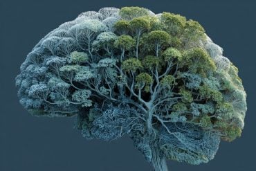
Summary: A new study has uncovered the brain circuits responsible for individual differences in how animals adapt to repeated visual threats. Using advanced neural recording and manipulation tools, researchers identified two distinct pathways in the brain that drive either persistent escape or rapid habituation in mice.
These differences are shaped by internal arousal states and beta oscillations in the amygdala, suggesting a neural basis for why some individuals remain fearful while others quickly adapt. The findings deepen our understanding of fear plasticity and may inform treatments for anxiety-related disorders like PTSD.
Key Facts:
- Two Escape Types: Mice show either sustained escape (T1) or rapid habituation (T2) to visual threats.
- Distinct Brain Circuits: T1 involves SC–VTA–BLA, while T2 follows the SC–MD–BLA circuit.
- Clinical Implications: Disrupted fear circuitry may underlie anxiety, phobias, and PTSD.
Source: SIAT
In a study published in Neuron, a research team led by Prof. WANG Liping from the Shenzhen Institutes of Advanced Technology (SIAT) of the Chinese Academy of Sciences revealed the neural circuit underlying individual differences in visual escape habituation.
Emotional responses, such as fear behaviors, are evolutionarily conserved mechanisms that enable organisms to detect and avoid danger, ensuring survival.

Since Darwin’s On the Origin of Species (1859) proposed that individual differences drive natural selection, understanding behavioral adaptation has become essential for unraveling biodiversity and survival strategies.
Repeated exposure to predators can elicit divergent coping strategies—habituation or sensitization—that are dependent on sensory inputs, internal physiological states, and prior experiences.
However, the neural circuits underlying individual variability in the regulation of internal states and habituation to repeated threats remain poorly understood.
To address this question, Prof. WANG’s team employed advanced techniques such as in vivo multichannel recording, fiber photometry, pupillometry and optogenetic manipulation to investigate how individual differences in arousal and internal states influence visual escape habituation.
Researchers found that distinct subcortical pathways from the superior colliculus to the amygdala and insula cortical pathways that govern two visual escape behaviors in two groups of mice.
They identified two distinct defensive behaviors-sustained rapid escape (T1) and rapid habituation (T2).
T1 involves superior colliculus (SC)/insular cortex-ventral tegmental area (VTA)-basolateral amygdala (BLA) pathway, whereas T2 relies on SC/insula -dorsomedial thalamus (MD)-BLA circuit. The MD integrates inputs from the SC and insula to regulate arousal and fear responses, while beta oscillations in BLA modulate fear states.
“Dysregulation of innate fear circuits is closely linked to many mental health conditions, including phobias, anxiety, and post-traumatic stress disorder (PTSD).
“Elucidating the neural circuitry underlying innate fear not only enhances our understanding of emotional disorders but also provides promising therapeutic targets for clinical interventions,” said Prof. WANG.
By elucidating the neural basis of individual differences in fear plasticity, this study highlights the central role of brain states in stress adaptation.
“Our work provides new insights into arousal modulation, internal states, and adaptive responses to visual threats,” said Prof. LIU Xuemei, one of the first authors of the study.
About this fear and neuroscience research news
Author: LU Qun
Source: SIAT
Contact: LU Qun – SIAT
Image: The image is credited to Neuroscience News
Original Research: Open access.
“Neural circuit underlying individual differences in visual escape habituation” by WANG Liping et al. Neuron
Abstract
Neural circuit underlying individual differences in visual escape habituation
Emotions like fear help organisms respond to threats.
Repeated predator exposure leads to adaptive responses with unclear neural mechanisms behind individual variability.
We identify two escape behaviors in mice—persistent escape (T1) and rapid habituation (T2)—linked to unique arousal states under repetitive looming stimuli.
Combining multichannel recording, circuit mapping, optogenetics, and behavioral analyses, we find parallel pathways from the superior colliculus (SC) to the basolateral amygdala (BLA) via the ventral tegmental area (VTA) for T1 and via the mediodorsal thalamus (MD) for T2. T1 involves heightened arousal, while T2 features rapid habituation.
The MD integrates SC and insular cortex inputs to modulate arousal and defensive behaviors.
This work reveals neural circuits underpinning adaptive threat responses and individual variability.


