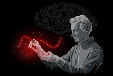
Summary: Responsive neurostimulation can remodel neural networks, leaving the brain less susceptible to epileptic seizures.
Source: University of Pittsburgh
Responsive neurostimulation (RNS) treats epilepsy by detecting seizures and intervening with a jolt of electric current. Over time, most patients find their seizures become fewer and further between. Now, for the first time, researchers at the University of Pittsburgh School of Medicine and UPMC have a better understanding of why this happens.
As reported today in JAMA Neurology, new evidence suggests RNS can remodel the brain to be less susceptible to seizures. Using patients’ brain signatures as a guide, the researchers hope to rapidly optimize the use of neurostimulation to help more people achieve seizure reduction.
“Right now, in epilepsy patients treated with responsive neurostimulation, you just have to wait and see whether their seizure frequency goes down,” said Mark Richardson, M.D., Ph.D., associate professor of neurological surgery at Pitt’s School of Medicine and director of epilepsy and movement disorders surgery at UPMC. “We don’t have a great way to predict who will respond. But there’s so much more data recorded on these RNS devices than we currently have the ability to analyze.”
In patients implanted with the RNS system, baseline brain activity is recorded for a month to characterize a person’s individual seizure patterns. This information is then used to train the stimulator to automatically respond to a seizure as it’s happening.
What no one has done before, Richardson said, is to analyze each individual recording of seizure activity captured by the device over time to see what is different about the brain activity of patients who experience a reduction in seizure frequency as a result of neurostimulation.
“I was expecting to find what was traditionally assumed–that the electric pulse acutely stops the seizure,” said lead author Vasileios Kokkinos, Ph.D., a neurosurgery research instructor at Pitt. “I realized this was not what was happening. I saw some examples of acute stopping of the seizures, but that only accounted for 5% of all seizures. It couldn’t explain the 60-70% of patients who responded to treatment.”
The researchers’ theory is that stimulation changes brain networks–the web of linkages between neurons–so that electrical rumblings at the neural epicenter can’t spread into a full-blown seizure.
“In epilepsy, brain networks get recruited to fire hyper-synchronously,” Richardson said. “What we think we’re doing when we stimulate is to desynchronize, over time, making it harder for the whole seizure network to come online.”
To test that hypothesis, Kokkinos and other members of Richardson’s Brain Modulation Laboratory compiled electrical brain activity data recorded on the RNS system in 11 patients with focal epilepsy treated at the University of Pittsburgh Comprehensive Epilepsy Center (UPCEC), an accredited level 4 epilepsy center. These patients started RNS therapy between 2015 and 2017 and were followed for up to two years.
Kokkinos found that people who ultimately experienced fewer seizures after starting neurostimulation treatment showed progressive reductions in spontaneous hyper-synchronous brain activity, starting as early as two months after the stimulator was first turned on. Richardson contrasted the faster, objective onset of these brain activity changes against the slower, subjective process of waiting to see symptom improvement in a patient’s self-reported diary of seizure activity.
“This study justifies my inherent enthusiasm for the RNS’s diagnostic potential beyond its proven therapeutic potential,” said Anto Bagi?, M.D., Ph.D., chief of UPMC’s Epilepsy Division, director of the UPCEC and professor of neurology at Pitt’s School of Medicine. He was not part of the study.
“It appears increasingly likely that this exquisite way of monitoring brain activity through a therapeutic tool will enable us to assess even a response to seizure medications without prolonged waiting and suffering for patients.”

Richardson hopes this kind of brain activity analysis will provide quicker feedback during the trial-and-error process of parameter adjustment, so that patients can see the long-term benefits of responsive neurostimulation sooner.
“Our next move is to incorporate what we’ve learned into a formal pipeline for analysis that might allow us in the future to predict which patients are responding before they’re able to report that to us clinically,” Richardson said.
Funding: This research was funded by the Walter L. Copeland Fund of the Pittsburgh Foundation and National Institutes of Neurological Disorders and Stroke grant R01 NS110424.
Additional authors on the study include Nathaniel Sisterson and Thomas Wozny, M.D., both from Pitt.
Source:
University of Pittsburgh
Media Contacts:
Erin Hare – University of Pittsburgh
Image Source:
The image is in the public domain.
Original Research: Closed access
Kokkinos V, Sisterson ND, Wozny TA, Richardson RM. “Association of Closed-Loop Brain Stimulation Neurophysiological Features With Seizure Control Among Patients With Focal Epilepsy”. JAMA Neurology. Published online April 15, 2019 doi:10.1001/jamaneurol.2019.0658
Abstract
Association of Closed-Loop Brain Stimulation Neurophysiological Features With Seizure Control Among Patients With Focal Epilepsy
Importance A bidirectional brain-computer interface that performs neurostimulation has been shown to improve seizure control in patients with refractory epilepsy, but the therapeutic mechanism is unknown.
Objective To investigate whether electrographic effects of responsive neurostimulation (RNS), identified in electrocorticographic (ECOG) recordings from the device, are associated with patient outcomes.
Design, Setting, and Participants Retrospective review of ECOG recordings and accompanying clinical meta-data from 11 consecutive patients with focal epilepsy who were implanted with a neurostimulation system between January 28, 2015, and June 6, 2017, with 22 to 112 weeks of follow-up. Recorded ECOG data were obtained from the manufacturer; additional system-generated meta-data, including recording and detection settings, were collected directly from the manufacturer’s management system using an in-house, custom-built platform. Electrographic seizure patterns were identified in RNS recordings and evaluated in the time-frequency domain, which was locked to the onset of the seizure pattern.
Main Outcomes and Measures Patterns of electrophysiological modulation were identified and then classified according to their latency of onset in relation to triggered stimulation events. Seizure control after RNS implantation was assessed by 3 main variables: mean frequency of seizure occurrence, estimated mean severity of seizures, and mean duration of seizures. Overall seizure outcomes were evaluated by the extended Personal Impact of Epilepsy Scale questionnaires, a patient-reported outcome measure of 3 domains (seizure characteristics, medication adverse effects, and quality of life), with a range of possible scores from 0 to 300 in which lower scores indicate worse status, and the Engel scale, which comprises 4 classes (I-IV) in which lower numbers indicate greater improvement.
Results Electrocorticographic data from 11 patients (8 female; mean [range] age, 35 [19-65] years; mean [range] duration of epilepsy, 19 [5-37] years) were analyzed. Two main categories of electrophysiological signatures of stimulation-induced modulation of the seizure network were discovered: direct and indirect effects. Direct effects included ictal inhibition and early frequency modulation but were not associated with improved clinical outcomes (odds ratio [OR], 0.67; 95% CI, 0.06-7.35; P > .99). Only indirect effects—those occurring remote from triggered stimulation—were associated with improved clinical outcomes (OR, infinity; 95% CI, –infinity to infinity; P = .02). These indirect effects included spontaneous ictal inhibition, frequency modulation, fragmentation, and ictal duration modulation.
Conclusions and Relevance These findings suggest that RNS effectiveness may be explained by long-term, stimulation-induced modulation of seizure network activity rather than by direct effects on each detected seizure.


