Summary: Researchers have developed new fluorescence sensors that are able to record dopamine activity in the living brain. The tool will help further understanding of dopamine’s role in reward and motivation.
Source: UC Davis.
UC Davis neuroscientist Lin Tian and her team, Tommaso Patriarchi, Gerard Broussard and Ruqiang Liang, have developed fluorescence sensors that are opening a new era for the optical recording of dopamine activity in the living brain.
The technology precisely captures where and when dopamine activity occurs in the brain within milliseconds and at the cellular level, producing a high-resolution map of dopamine transients associated with behaviors, such as learning.
A broad application of this tool will further understanding of dopamine activity underlying motivation, reward and movement, and pave the way to discover effective and novel therapeutics for depression, addiction and drug abuse.
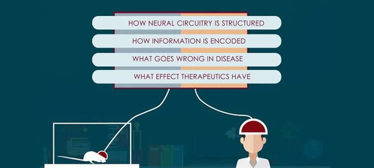
What is the current knowledge about the brain and its circuitry, and what don’t we know?
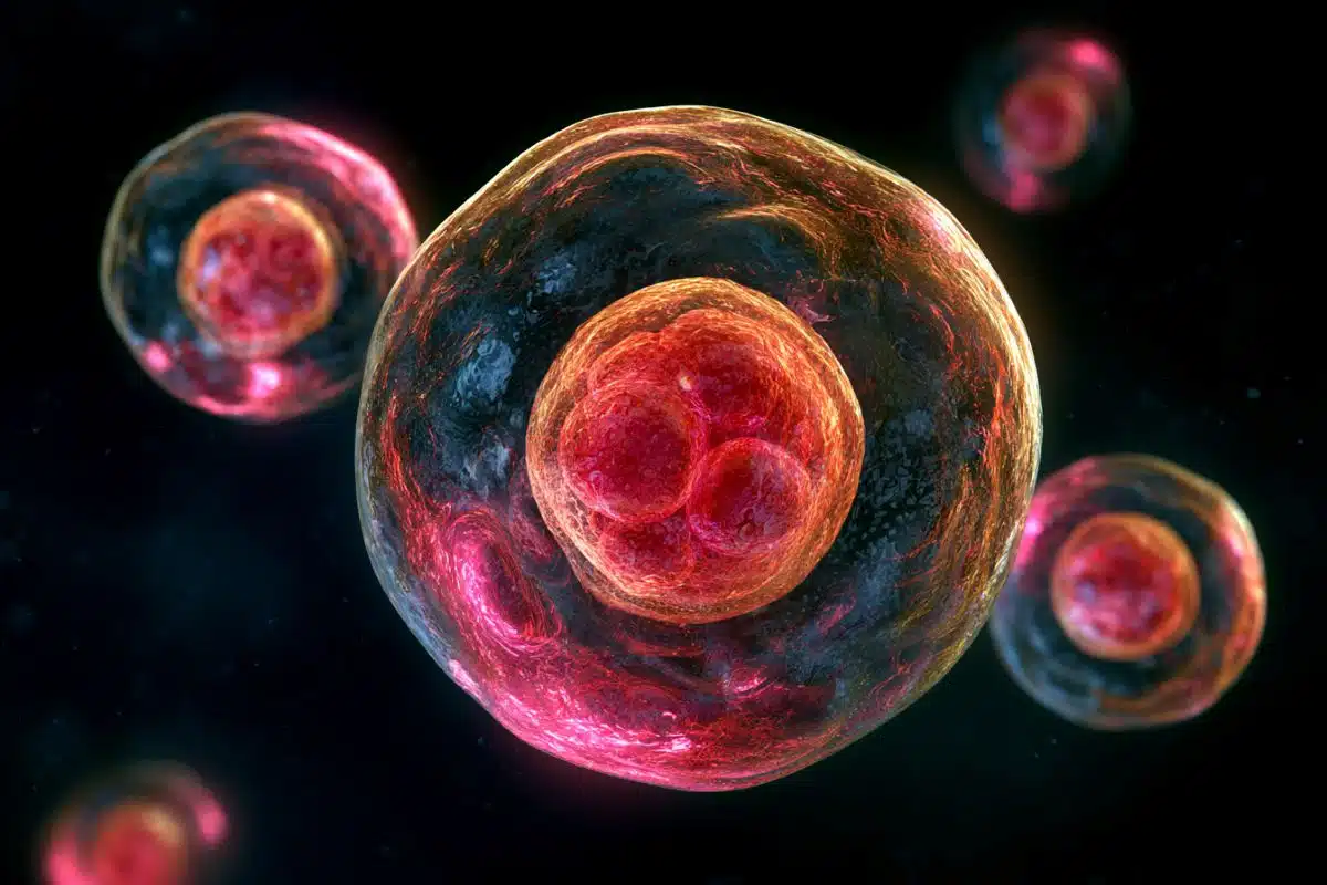
The brain’s “wiring” contains billions of neurons interconnected by trillions of synapses, the space between the end of a nerve cell and another cell that allows the transmission of impulses by chemicals called neurotransmitters. Electrophysiology and imaging studies have provided details on the structure and function of nerve cells and how an action potential at the synapse allows the transmission of signals that govern essential functions, from learning, memory and behavior to movement and sensations. But what we don’t understand as well is what is happening at the molecular level and which factors, conditions and chemical levels drive neurotransmitter release at the synapse.
Why is understanding brain circuitry important?
Altered dynamics of neurotransmitters and neuromodulators are associated with a number of neurological and psychiatric diseases, including Parkinson’s disease, schizophrenia and addiction. Hence, drugs that mimic or block neuromodulators have become important components in the treatment of these disorders. The neurotransmitter dopamine, for example, plays an important role in movement, attention, learning and the brain’s pleasure and reward system. Mental illnesses, such as depression, can occur when this process isn’t working correctly.
Although neuroscientists have discovered more than 100 neurotransmitters and neuromodulators and detailed their structure and function, we don’t know the precise mechanisms by which these molecules regulate the dynamics of healthy and diseased neural circuitry. Linking complex neural phenomena to the structure and function of their composite neural circuits requires a thorough understanding of patterns of neural activity, and the ability to relate this to physiological processes and behavior.
Would you summarize your study published in Science?
We report the development of the first class of fluorescent protein-based optical dopamine (DA) indicators, which we named dLight1, capable of directly reporting spatial and temporal release of DA with high-resolution both in vitro and in vivo. By labeling a specific neuronal population in living animals, we now can track the dynamic change of released dopamine that occurs within milliseconds and at cellular level during specific activities, such as running and reward learning. dLight overcomes major barriers in the field, and dramatically increases the resolution that we can measure dopamine in the brain of behaving mice. This technology is a game-changer. It can record spatioltemporal coding of dopamine in the brain (creating dopamine-transient map) in high resolution that codes motivation, movement, learning and drug abuse.
We can leverage these tools to develop sensors for other neuromodulators, including norepinephrine, serotonin, melatonin and opioid neuropeptides. This toolset offers the ability to gain vital information about release sites and kinetics for these endogenous ligands, thus open a new era for developing effective drugs for neurological disorders and revealing brain functions that underly motivation, movement, learning and drug abuse.
Why is this study important?
Our specially designed sensors combined with optical imaging techniques provides a powerful tool for analyzing and engineering functional neural circuits associated with learning, memory, behavior and disease states. We can leverage these tools to develop sensors for other neuromodulators, including norepinephrine, serotonin, melatonin and opioid neuropeptides and to empower the search for novel therapeutic treatments.
Funding: Funding for this study was provided by BRAIN Initiative, National Institute of Health, Catharina Foundation, Heritage Medical Research Institute.
Source: Carole Gan – UC Davis
Publisher: Organized by NeuroscienceNews.com.
Image Source: NeuroscienceNews.com image is credited to Laboratory of Lin Tian at UC Davis.
Original Research: Abstract for “Ultrafast neuronal imaging of dopamine dynamics with designed genetically encoded sensors” by Tommaso Patriarchi, Jounhong Ryan Cho, Katharina Merten, Mark W. Howe, Aaron Marley, Wei-Hong Xiong, Robert W. Folk, Gerard Joey Broussard, Ruqiang Liang, Min Jee Jang, Haining Zhong, Daniel Dombeck, Mark von Zastrow, Axel Nimmerjahn, Viviana Gradinaru, John T. Williams, and Lin Tian in Science. Published May 31 2018.
doi:10.1126/science.aat4422
[cbtabs][cbtab title=”MLA”]UC Davis “New Optical Probe Allows Ultrafast Imaging of Dopamine in the Brain.” NeuroscienceNews. NeuroscienceNews, 4 June 2018.
<https://neurosciencenews.com/dopamine-imaging-optical-probe-9239/>.[/cbtab][cbtab title=”APA”]UC Davis (2018, June 4). New Optical Probe Allows Ultrafast Imaging of Dopamine in the Brain. NeuroscienceNews. Retrieved June 4, 2018 from https://neurosciencenews.com/dopamine-imaging-optical-probe-9239/[/cbtab][cbtab title=”Chicago”]UC Davis “New Optical Probe Allows Ultrafast Imaging of Dopamine in the Brain.” https://neurosciencenews.com/dopamine-imaging-optical-probe-9239/ (accessed June 4, 2018).[/cbtab][/cbtabs]
Abstract
Ultrafast neuronal imaging of dopamine dynamics with designed genetically encoded sensors
Neuromodulatory systems exert profound influences on brain function. Understanding how these systems modify the operating mode of target circuits requires measuring spatiotemporally precise neuromodulator release. We developed dLight1, an intensity-based genetically encoded dopamine indicator, to enable optical recording of dopamine dynamics with high spatiotemporal resolution in behaving mice. We demonstrated the utility of dLight1 by imaging dopamine dynamics simultaneously with pharmacological manipulation, electrophysiological or optogenetic stimulation, and calcium imaging of local neuronal activity. dLight1 enabled chronic tracking of learning-induced changes in millisecond dopamine transients in striatum. Further, we used dLight1 to image spatially distinct, functionally heterogeneous dopamine transients relevant to learning and motor control in cortex. We also validated our sensor design platform for developing norepinephrine, serotonin, melatonin, and opioid neuropeptide indicators.