Summary: New research reveals how our brains coordinate with others during dancing, highlighting the importance of shared rhythm and visual contact. When dancers moved to the same song and could see each other, unique neural signals emerged to support social coordination.
Surprisingly, the brain responded most to subtle knee-bouncing movements—suggesting these actions may hold a special role in syncing people together. The findings offer broader insights into how we engage socially by aligning motion and sensory input, extending well beyond the dance floor.
Key Facts:
- Shared Rhythm & Vision: Social coordination signals appeared only when dancers heard the same song and saw each other.
- Bounce Sensitivity: The brain showed the strongest response to subtle knee-bouncing, despite its weak physical amplitude.
- Neural Separation: The study differentiated brain signals for self-movement, partner-following, and social coordination.
Source: SfN
Dancing fluidly with another involves social coordination. This skill entails aligning movements with others while also processing dynamic sensory information, like sounds and visuals.
In a new JNeurosci paper, Félix Bigand and Giacomo Novembre, from the Italian Institute of Technology, Rome, and colleagues report their findings on how the brain drives social coordination during dance.

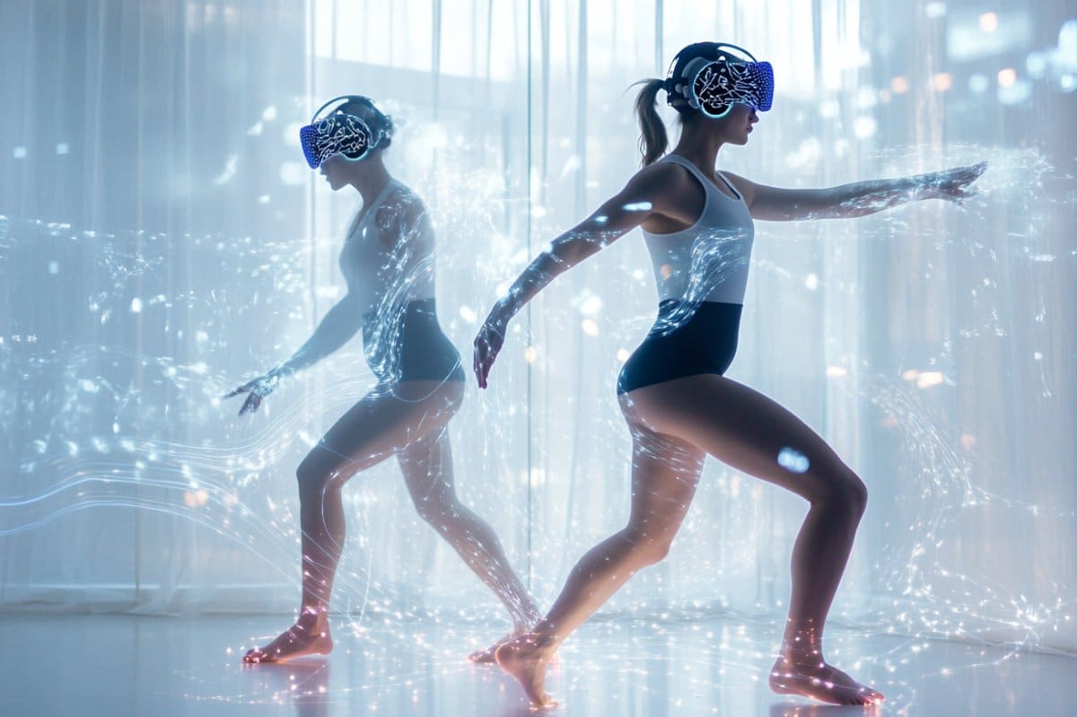
The researchers recruited pairs of inexperienced dancers and recorded their brain activity, whole-body movements, and muscle activity as they danced to the same or different songs.
The researchers also manipulated whether dancers could or could not see each other. These methods unveiled distinct neural signals for music processing, self-generated movements, movements generated by following a partner, and social coordination.
Neural signals for social coordination that enabled synchronized movements between people occurred only when dancers were moving to the same song and could see each other.
Says Bigand, “What was perhaps most peculiar was we found that out of the 15 different movements we recorded, the brain was most sensitive to bouncing or flexing of the knees [during social coordination].
“This was strange because bouncing had relatively weak amplitudes [or strength] compared to most of the other movements. For the brain to respond more to a weaker movement, like bounce, suggests it has a unique role in social coordination.”
According to the authors, this work advances our understanding of social interaction beyond dancing because it sheds light on how the brain supports socially engaging activities while integrating dynamic sensory information.
Bigand also emphasizes that the methods used to unravel distinct neural signals for different kinds of sensory information processing may improve the applicability of future preclinical work to reality.
About this social neuroscience research news
Author: SfN Media
Source: SfN
Contact: SfN Media – SfN
Image: The image is credited to Neuroscience News
Original Research: Closed access.
“EEG of the Dancing Brain: Decoding Sensory, Motor, and Social Processes During Dyadic Dance” by Félix Bigand et al. Journal of Neuroscience
Abstract
EEG of the Dancing Brain: Decoding Sensory, Motor, and Social Processes During Dyadic Dance
Real-world social cognition requires processing and adapting to multiple dynamic information streams. Interpreting neural activity in such ecological conditions remains a key challenge for neuroscience.
This study leverages advancements in de-noising techniques and multivariate modeling to extract interpretable EEG signals from pairs of participants (male-male, female-female, and male-female) engaged in spontaneous dyadic dance.
Using multivariate temporal response functions (mTRFs), we investigated how music acoustics, self-generated kinematics, other-generated kinematics, and social coordination uniquely contributed to EEG activity. Electromyogram recordings from ocular, face, and neck muscles were also modeled to control for artifacts.
The mTRFs effectively disentangled neural signals associated with four processes: (I) auditory tracking of music, (II) control of self-generated movements, (III) visual monitoring of partner movements, and (IV) visual tracking of social coordination.
We show that the first three neural signals are driven by event-related potentials: the P50-N100-P200 triggered by acoustic events, the central lateralized movement-related cortical potentials triggered by movement initiation, and the occipital N170 triggered by movement observation.
Notably, the (previously unknown) neural marker of social coordination encodes the spatiotemporal alignment between dancers, surpassing the encoding of self- or partner-related kinematics taken alone.
This marker emerges when partners can see each other, exhibits a topographical distribution over occipital areas, and is specifically driven by movement observation rather than initiation. Using data-driven kinematic decomposition, we further show that vertical bounce movements best drive observers’ EEG activity.
These findings highlight the potential of real-world neuroimaging, combined with multivariate modeling, to uncover the mechanisms underlying complex yet natural social behaviors.


