

Summary: Researchers have mapped four new areas of the human anterior prefrontal cortex that play critical roles in cognitive processing. Two of these newly mapped areas are larger in females than in males.
Source: Human Brain Project
Researchers of the Human Brain Project (HBP) have mapped four new areas of the human anterior prefrontal cortex that plays a major role in cognitive functions. Two of the newly identified areas are relatively larger in females than in males.
The human dorsolateral prefrontal cortex is involved in cognitive control including attention selection, working memory, decision making and planning of actions. Changes in this brain region are suspected to play a role in schizophrenia, obsessive-compulsive disorder, depression and bipolar disorder, making it an important research target.
Researchers from Forschungszentrum Jülich and Heinrich-Heine University Düsseldorf now provide detailed, three-dimensional maps of four new areas of the brain region.
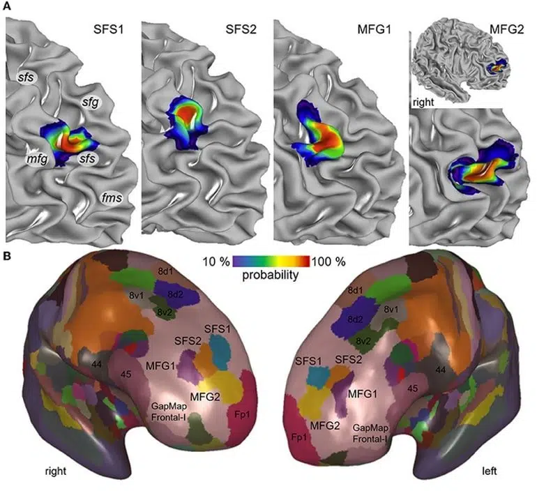
In order to identify the borders between brain areas, the researchers statistically analysed the distribution of cells (the cytoarchitecture) in 10 post mortem human brains. After reconstructing the mapped areas in 3D, the researchers superimposed the maps of the 10 different brains and generated probability maps that reflect how much the localization and size of each area varies among individuals.
High inter-subject variability has been a major challenge for prior attempts to map this brain region leading to considerable discrepancies in pre-existing maps and inconclusive information making it very difficult to understand the specific involvement of individual brain areas in the different cognitive functions.
The new probabilistic maps account for the variability between individuals and can be directly superimposed with datasets from functional studies in order to directly correlate structure and function of the areas.
When comparing the brains of female and male tissue donors, the researchers found that the relative volumes of two of the newly identified areas were significantly larger in female than in male brains. This finding may be related to sex differences in cognitive function and behaviour as well as in the prevalence and symptoms of associated brain diseases.
The maps are being integrated into the Julich Brain Atlas that is openly accessible via EBRAINS.
About this brain mapping research news
Author: Peter Zekert
Source: Human Brain Project
Contact: Peter Zekert – Human Brain Project
Image: The image is credited to Bruno et al
Original Research: Open access.
“Cytoarchitecture, intersubject variability, and 3D mapping of four new areas of the human anterior prefrontal cortex” by Bruno et al. Frontiers in Neuroanatomy
Abstract
Cytoarchitecture, intersubject variability, and 3D mapping of four new areas of the human anterior prefrontal cortex
The dorsolateral prefrontal cortex (DLPFC) plays a key role in cognitive control and executive functions, including working memory, attention, value encoding, decision making, monitoring, and controlling behavioral strategies. However, the relationships between this variety of functions and the underlying cortical areas, which specifically contribute to these functions, are not yet well-understood.
Existing microstructural maps differ in the number, localization, and extent of areas of the DLPFC. Moreover, there is a considerable intersubject variability both in the sulcal pattern and in the microstructure of this region, which impedes comparison with functional neuroimaging studies.
The aim of this study was to provide microstructural, cytoarchitectonic maps of the human anterior DLPFC in 3D space. Therefore, we analyzed 10 human post-mortem brains and mapped their borders using a well-established approach based on statistical image analysis. Four new areas (i.e., SFS1, SFS2, MFG1, and MFG2) were identified in serial, cell-body stained brain sections that occupy the anterior superior frontal sulcus and middle frontal gyrus, i.e., a region corresponding to parts of Brodmann areas 9 and 46.
Differences between areas in cytoarchitecture were captured using gray level index profiles, reflecting changes in the volume fraction of cell bodies from the surface of the brain to the cortex-white matter border.
A hierarchical cluster analysis of these profiles indicated that areas of the anterior DLPFC displayed higher cytoarchitectonic similarity between each other than to areas of the neighboring frontal pole (areas Fp1 and Fp2), Broca’s region (areas 44 and 45) of the ventral prefrontal cortex, and posterior DLPFC areas (8d1, 8d2, 8v1, and 8v2).
Area-specific, cytoarchitectonic differences were found between the brains of males and females. The individual areas were 3D-reconstructed, and probability maps were created in the MNI Colin27 and ICBM152casym reference spaces to take the variability of areas in stereotaxic space into account.
The new maps contribute to Julich-Brain and are publicly available as a resource for studying neuroimaging data, helping to clarify the functional and organizational principles of the human prefrontal cortex.


