Summary: Researchers have developed DELTA, a powerful new imaging method that maps brain-wide synaptic changes during learning. By labeling synaptic proteins before and after behavioral training, scientists can now visualize where and how neural connections shift over time.
The technique revealed changes in the synaptic protein GluA2 in specific brain regions linked to new learning and across the brain in enriched environments. DELTA opens the door to uncovering the molecular basis of learning and memory by bridging the gap between behavior and cellular changes.
Key Facts:
- Whole-Brain View: DELTA maps where synaptic protein changes occur during learning.
- Learning in Action: Protein GluA2 changes reveal brain regions engaged by new tasks.
- Versatile Tool: DELTA can track synaptic shifts in both standard and enriched environments.
Source: HHMI
Janelia researchers have developed a new way to map how individual connections between neurons change across the entire brain during learning, giving scientists a new view into how changes in behavior show up in the brain.
Neurons communicate with each other by passing millisecond-long signals across tiny junctions called synapses. Experiences cause the strength of these connections to change – a process called synaptic plasticity, which underlies learning and memory. But exactly where in the brain these changes occur during the learning process is largely unknown.

Janelia researchers have devised a new way to see where these synaptic changes are happening across the entire brain. The work was led by the Spruston and Lavis labs and the lab of former Janelia Senior Group Leader Karel Svoboda.
The new imaging method, DELTA, provides scientists with a brain-wide map of how individual synaptic proteins change over time. Proteins important for synaptic plasticity are known to be degraded or synthesized as synaptic connections change. So, tracking these synaptic protein changes during learning enables scientists to also understand how synaptic connections change.
This information allows researchers to home in on areas of the brain that might be important for learning and memory and helps them figure out the molecular mechanisms behind the synaptic changes.
“One of the problems in figuring out which molecules are in charge of what changes is that it’s very hard to know where those changes are happening. We didn’t have good way of figuring out where in the brain things are changing so we can focus our attention on the most interesting parts,” says Boaz Mohar, a Research Scientist in the Spruston Lab who led the research.
“Now we have a different way of looking at our manipulations and how they affect the circuit at the brain-wide level by using imaging to drill down on the subcellular structure. This helps us bridge that gap between behavior and mechanism.”
The method works by first labeling a synaptic protein of interest in the mouse brain with a bright Janelia Fluor (JF) dye. To investigate how synaptic connections change during learning, the researchers used mice that had already been trained to on a simple task: learning to associate two distinct visual cues with a water reward.
After this initial labeling, the mice were split into two groups: one group received water randomly at the two cues, as they had prior to labeling, while the task for the other group was modified such that only one of the cues was associated with the water reward. After a few days, once the researchers saw a clear difference between the two groups’ actions, the same synaptic protein of interest was labeled with a different JF dye in both groups of mice.
Over those days, some of the original labeled proteins are degraded and some new proteins are synthesized, incorporating the second dye. By imaging the entire brain, researchers can see where the synaptic proteins have changed in both groups of mice, giving them a hint as to where changes in synaptic connections are happening during learning.
The researchers saw that learning the modified task caused the synaptic protein they studied, GluA2, to change in specific brain regions.
The researchers also used the new method to see differences in protein turnover in mice living in a normal environment and mice living in an enriched environment with toys and companions. In this case, putting a mouse in an enriched environment caused widespread changes in GluA2 across the entire brain.
By enabling researchers to see these brain-wide changes, DELTA gives scientists a starting point for follow up studies to detail the cellular and molecular mechanisms behind learning and memory.
As a next step, the researchers are working to add additional features to DELTA that will allow them to pinpoint when proteins are changing during the few days the animals are learning a task.
One of the lab heads who led the project, Nelson Spruston, says the development of DELTA exemplifies the collaborative spirit of the research campus, with scientists across Janelia lending their knowledge in chemistry, imaging, behavior, and genetics to the project. The team also collaborated with experts outside Janelia, including researchers at Northwestern University who helped to validate the method.
Now, the team is working with scientists around the world to help them use DELTA to track synaptic changes in their own research, collaborations enabled through Janelia’s Visiting Scientist Program.
“This is a quintessential Janelia project,” Spruston says. “It involved doing something that nobody has ever done before and required collaboration with lots of people who contributed expertise to make this all possible.”
About this brain mapping and synaptic plasticity research news
Author: Nanci Bompey
Source: HHMI
Contact: Nanci Bompey – HHMI
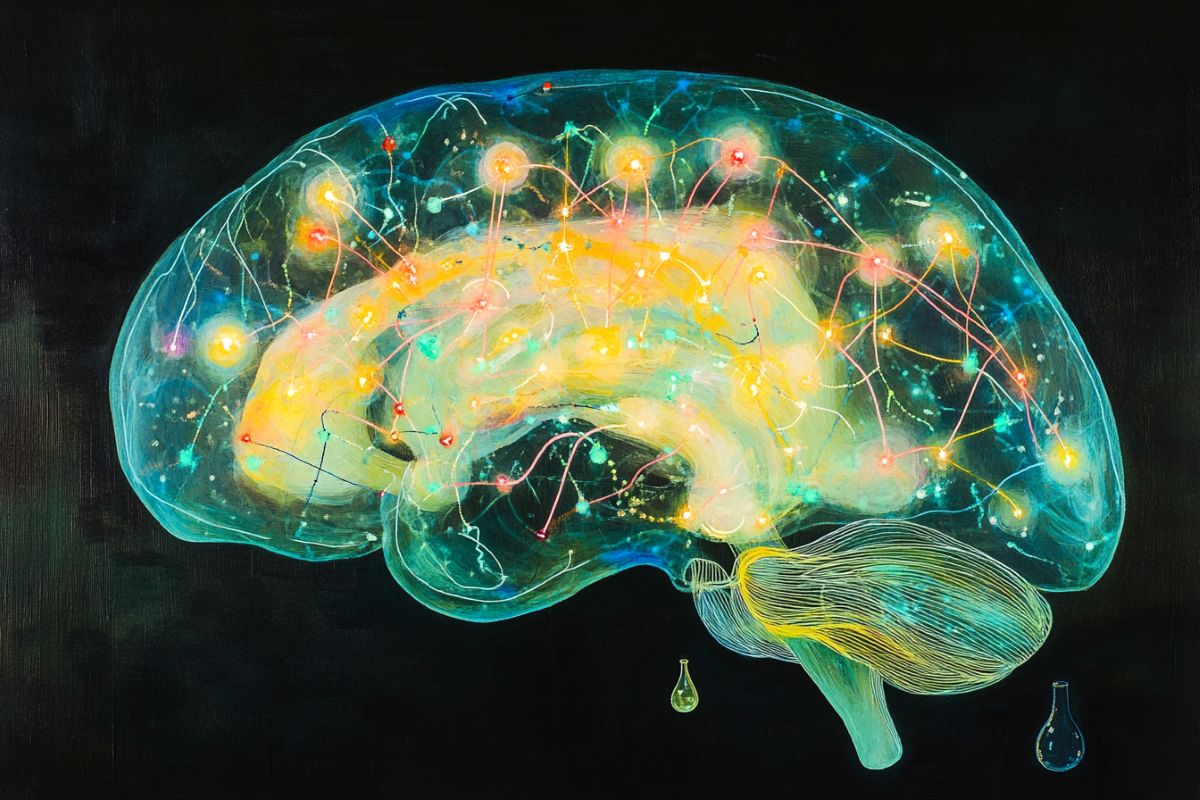
Image: The image is credited to Neuroscience News
Original Research: Open access.
“DELTA: a method for brain-wide measurement of synaptic protein turnover reveals localized plasticity during learning” by Boaz Mohar et al. Nature Neuroscience
Abstract
DELTA: a method for brain-wide measurement of synaptic protein turnover reveals localized plasticity during learning
Synaptic plasticity alters neuronal connections in response to experience, which is thought to underlie learning and memory. However, the loci of learning-related synaptic plasticity, and the degree to which plasticity is localized or distributed, remain largely unknown.
Here we describe a new method, DELTA, for mapping brain-wide changes in synaptic protein turnover with single-synapse resolution, based on Janelia Fluor dyes and HaloTag knock-in mice.
During associative learning, the turnover of the ionotropic glutamate receptor subunit GluA2, an indicator of synaptic plasticity, was enhanced in several brain regions, most markedly hippocampal area CA1. More broadly distributed increases in the turnover of synaptic proteins were observed in response to environmental enrichment.
In CA1, GluA2 stability was regulated in an input-specific manner, with more turnover in layers containing input from CA3 compared to entorhinal cortex. DELTA will facilitate exploration of the molecular and circuit basis of learning and memory and other forms of plasticity at scales ranging from single synapses to the entire brain.


