

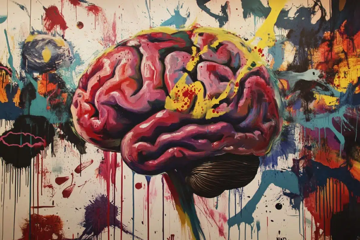
Summary: New research has uncovered that brain signals for aggression in male mice and sexual arousal in female mice are encoded by similar neural mechanisms. The studies found that a specific type of neural signal, called a line attractor, represents the intensity and persistence of these emotional states.
In the case of aggression, this signal builds up over time and slowly decays after the stimulus is removed, resembling how humans calm down after anger. The findings suggest that different emotions may share common neural pathways, potentially offering insights into mental health treatments.
Key Facts:
- Aggression and arousal in mice are both encoded by a neural “line attractor.”
- These signals build up and persist over time, similar to human emotional states.
- Findings may inform future treatments for emotional and mental health conditions.
Source: CalTech
A series of three papers from neuroscientist David J. Anderson’s laboratory, two in the journal Nature and one in the journal Cell, reveal new insights into the neural signals underlying internal emotional states including aggression and sexual arousal.
The studies show that the state of aggression in male mice and the state of arousal in female mice are both encoded by a common type of signal in the brain.
The shape of emotions
These findings are the result of collaborations within the group of Anderson, who is the Seymour Benzer Professor of Biology, Howard Hughes Medical Institute Investigator, Tianqiao and Chrissy Chen Leadership Chair, and director of the Tianqiao and Chrissy Chen Institute for Neuroscience at Caltech.
The three new studies all build upon previous research from the Anderson lab, in particular a study published in Cell last year led by Computation and Neural Systems graduate student Aditya Nair, and former postdoctoral scholar Ann Kennedy, who is now an associate professor at the Scripps Research Institute.
In that study, researchers discovered a neural signal encoding the persistence and intensity of an internal state of aggression. Nair, who is also a co-first author on the two recent Nature papers and second author on the new Cell paper, used machine learning to model brain activity, which revealed that the neural signal causing the state of aggression in mice was a line attractor.
A line attractor is a specific pattern of activity, created by the interconnections between brain cells, that follows the shape of a valley. In a graph showing the flow of energy among neurons over time, the energy in a line attractor system tends to flow down the valley, like a ball rolling down into a trough. Once neural energy has reached the bottom, it tends to stay there and flow along a line, like a river moving along the bottom of a valley.
In the line attractor signal encoding aggression, the farther that neural energy flows along the line, the more the animal’s aggressive state escalates. Then after a fight, it takes time for the neural energy to flow back out of the valley. The researchers speculate that this gradual decay may correspond to the time it takes someone to calm down if they are very upset or angry.
Nair says this finding was unexpected because, while line attractors had been observed in the cortex and hippocampus (which are evolutionarily recent brain regions that control higher cognitive functions), many had assumed that the hypothalamus (an evolutionarily ancient region that controls instinctive behaviors) would not possess these types of signals.
The next challenges, says Nair, were “to really test our theory and see if this signal was indeed a property of the brain networks we were directly observing, to understand what mechanisms sustain this network, and to ask if this was unique to aggression or if it reflected a common principle for the brain to represent emotion states.”
The proof is in the hypothalamus
Because the original study was based on machine learning modeling, the researchers did not yet have evidence that the signal of escalating aggressiveness was encoded by local neural circuits.
While the researchers were observing activity in a specific region of the hypothalamus, it was possible the line attractor signal was being created in another brain region and passed along to the hypothalamus via long-range connections.
To answer that question, postdoctoral scholar Amit Vinograd and Nair performed a technically challenging “record and play back” type of experiment designed to test whether the neural signal could be reproduced in a laboratory setting by reactivating the right cells in mice brains.
This required that the researchers use sophisticated techniques to observe neural activity in the hypothalamus in real time while a mouse was in an aggressive state, identify which neurons contributed to the aggressiveness line attractor, and then restimulate those specific neurons to see whether the line attractor could be reproduced.
In other words, if an external stimulus caused the neural energy to flow into the valley, would directly stimulating the observed neurons push the system into the same valley as well? If so, it would imply that the line attractor could be generated locally by the neurons being observed and was not passively “inherited” from some other brain region.
“This experiment was one of those rare ‘movie moments’ you don’t get often in science and, to me, really showed the power of collaboration,” Nair says.
While the researchers used machine learning to model the neural data and locate the line attractor in the brain, they created a hologram of the specific neurons they needed to activate. Then, they used a laser to reactivate those neurons.
When the researchers artificially reactivated the individual neurons that were making up the line attractor signal, they observed that the cells “integrated” the activation inputs, gradually accumulating the new neural signals into a stronger, more persistent signal, which was the line attractor.
This accumulation could be compared to more water flowing into a river and pushing the river farther along the bottom of a valley.
“Once we saw that the signal was actually intrinsic in the network, we wanted to know how it was being created,” Vinograd says.
“So, then we did experiments to test the functional connectivity of neurons by activating single cells to see whether other cells in the network also light up. We saw what we call ‘recurrent connectivity,’ which means that a specific cell population—those that make up the line attractor—are interconnected to one another.
“This interconnectivity allows them to do this computation that integrates the information; the strength of connectivity increases because the cells are amplifying each other.”
This amplification is what creates the tendency of the energy to flow into the line attractor shape. The study marked the first direct experimental evidence of line attractor dynamics in a mammalian brain—something that was previously only theorized to exist.
The results are described in the paper “Causal evidence of a line attractor encoding an affective state,” published in Nature.
Neuropeptides facilitate the signal of aggressiveness
In another study published in Cell, researchers in the Anderson lab, led by postdoctoral scholar George Mountoufaris, found that the neuropeptides oxytocin and vasopressin—brain chemicals important in social behavior and social learning—are necessary for the aggressiveness line attractor to form.
Building again upon prior studies from the lab, the researchers hypothesized that the aggressiveness signal, which persists for relatively longer than other brain signals, is implemented in neural circuits with the use of slow-acting chemical messengers including neuropeptides.
While many neural signals are implemented with the more common and fast-acting chemical messenger glutamate, neuropeptides like oxytocin and vasopressin can influence the activity of neural circuits with longer-lasting and wider-reaching effects in the brain.
To explore the role of these neuropeptides in implementing the signal of aggressiveness, Mountoufaris invented a new technique called “CRISPRoscopy,” which combines the gene-editing technology CRISPR with single-cell calcium imaging methods to record brain activity.
Using the technique, researchers disrupted specific neurons’ ability to detect oxytocin and vasopressin. They then observed the effect of this genetic disruption on both the animals’ social behavior and on their brain activity.
“While aggression was not completely abolished, the animals exhibited reduced aggression and fought with less vigor,” Mountoufaris says. “Without the signaling of oxytocin and vasopressin, the mice were unable to sustain the anger state required to escalate the aggression.”
Inside the brain, the disruption affected how long individual neurons, as well as populations of neurons, remained active once triggered by an aggression-promoting stimulus.
“The persistence of neural responses at the single-cell level was significantly diminished,” Mountoufaris says. “And the population-level activity was completely compromised.”
These findings showed that the line attractor did not form without the neuropeptide signaling, indicating that oxytocin and vasopressin play a critical role in the implementation of the aggressiveness signal.
Mountoufaris says this result was surprising and significant because most previous theoretical studies had discounted the role of such slow chemical messengers in generating line attractors.
The full study, “A line attractor encoding a persistent internal state requires neuropeptide signaling,” is published in Cell.
A neural signal of sexual arousal
While the hypothalamus in male mice has a line attractor encoding a signal of anger, a new study led by graduate alum Mengyu Liu (PhD ’24) and Nair found that the same region in female mice has a line attractor encoding sexual receptivity, which may generate an internal state of sexual arousal.
In a previous study led by Liu, researchers discovered a female-specific type of neuron within the hypothalamus that promotes sexual receptivity. Surprisingly, however, as females proceeded through their estrus cycle (which in mice lasts about a week), there was no change in the overall level of activity of these neurons whether the females were in their receptive or non-receptive phase.
To better understand the activity of the sexual receptivity-promoting cell population, Nair applied the same machine learning techniques to the female mouse data.
“We think of sexual arousal as an internal state, which has the same qualities as an aggressive state: persistence and intensity,” Nair says. “This gave us to opportunity to really ask if the line attractor we observed in aggression was specific to that state or was a common type of brain computation that is reused for different types of emotion states, including sexual arousal.”
This approach indeed revealed a line attractor encoding the signal of sexual receptivity in females. During the “receptive” stages of the estrus cycle, the arousal signal moved along the bottom of the valley in response to repeated sexual contact by male mice.
As male mice sniffed, mounted, and mated with female mice, the female mouse brains accumulated those inputs, and they were more likely to display receptive behavior in response to males.
“One surprising discovery is the gradual ramping up of sexual arousal in females during mating, which can occur over several minutes or even longer,” Liu says.
The authors speculate that this ramping and persistent neural activity (which flows in a similar pattern to the aggressiveness signal in males) may serve to keep the females “interested” in the male in between bouts of mounting, which are intermittent and sporadic.
The study also revealed that the mating line attractor was only observed during specific phases of the estrus cycle, when the females were sexually receptive. If females were mounted by males during the non-receptive phase of their cycle, the line attractor was not observed.
“We found that achieving this heightened state of arousal depends on being in the appropriate hormonal state,” says Liu.
“Without the right hormonal conditions, even repeated successful copulations—essentially nonconsensual encounters—may not lead to sexual arousal in the female.”
“Historically, scientific research has disproportionately focused on males, leaving female-specific biological questions largely underexplored,” Liu says.
“There are significant differences between the sexes in physiology and brain function, which limit women’s ability to fully benefit from male-centered scientific findings. Addressing this gap by prioritizing female subjects and questions is critical to advancing gender-inclusive science and improving women’s health.”
Liu says the findings of the study can ultimately guide research to inform health care providers and the public about the natural fluctuations in a woman’s emotional state during sexual experiences due to hormonal changes and may also provide justification for the development of hormone therapies aimed at improving sexual health in women.
The study is called “Encoding of female mating dynamics by a hypothalamic line attractor” and is published in Nature.
A shared language for encoding emotions
The three new studies reveal unique findings about how internal states of male aggressiveness and female sexual receptivity are implemented in the brain. The authors say these internal states likely exist in humans as well, where we may consciously perceive them as feelings that we call “anger” and “sexual arousal,” respectively.
Altogether, the studies suggest that the persistence and intensity of emotional states might be encoded by a common property of certain neural networks in the brain in the form of line attractors.
Because attractors are stable states of brain activity that are difficult to dispel once they form, it has been hypothesized that they may underlie certain types of long-lasting mental illness, such as depression.
“We’re very excited about what these findings could mean for mental health conditions,” Nair says. “All these studies use cutting-edge tools, which, when combined with machine learning, can unveil both how attractors might change with different disorders and how we might be able to intervene with mental health therapies.”
“These studies have opened up a new era of work in my laboratory using computational approaches,” says Anderson. While these approaches are a mainstay of research in other fields at Caltech, Anderson had not previously applied them to his own work.
“In the best tradition of Caltech,” Anderson says, “it was the expertise of a graduate student that brought this new approach to our research, with very exciting results and implications.”
Funding:
Funding for this research was provided by the National Institutes of Health (NIH) BRAIN Initiative, the Howard Hughes Medical Institute (HHMI), and the Agency for Science, Technology and Research of Singapore (A*STAR).
About this aggression, arousal, and neuroscience research news
Author: David J. Anderson
Source: CalTech
Contact: David J. Anderson – CalTech
Image: The image is credited to Neuroscience News
Original Research: Open access.
“Causal evidence of a line attractor encoding an affective state” by David J. Anderson et al. Nature
Open access.
“Encoding of female mating dynamics by a hypothalamic line attractor” by David J. Anderson et al. Nature
Open access.
“A line attractor encoding a persistent internal state requires neuropeptide signaling” by David J. Anderson et al. Cell
Abstract
Causal evidence of a line attractor encoding an affective state
Continuous attractors are an emergent property of neural population dynamics that have been hypothesized to encode continuous variables such as head direction and eye position.
In mammals, direct evidence of neural implementation of a continuous attractor has been hindered by the challenge of targeting perturbations to specific neurons within contributing ensembles. Dynamical systems modelling has revealed that neurons in the hypothalamus exhibit approximate line-attractor dynamics in male mice during aggressive encounters.
We have previously hypothesized that these dynamics may encode the variable intensity and persistence of an aggressive internal state. Here we report that these neurons also showed line-attractor dynamics in head-fixed mice observing aggression. This allowed us to identify and manipulate line-attractor-contributing neurons using two-photon calcium imaging and holographic optogenetic perturbations.
On-manifold perturbations yielded integration of optogenetic stimulation pulses and persistent activity that drove the system along the line attractor, while transient off-manifold perturbations were followed by rapid relaxation back into the attractor. Furthermore, single-cell stimulation and imaging revealed selective functional connectivity among attractor-contributing neurons.
Notably, individual differences among mice in line-attractor stability were correlated with the degree of functional connectivity among attractor-contributing neurons. Mechanistic recurrent neural network modelling indicated that dense subnetwork connectivity and slow neurotransmission best recapitulate our empirical findings.
Our work bridges circuit and manifold levels, providing causal evidence of continuous attractor dynamics encoding an affective internal state in the mammalian hypothalamus.
Abstract
Encoding of female mating dynamics by a hypothalamic line attractor
Females exhibit complex, dynamic behaviours during mating with variable sexual receptivity depending on hormonal status. However, how their brains encode the dynamics of mating and receptivity remains largely unknown.
The ventromedial hypothalamus, ventrolateral subdivision contains oestrogen receptor type 1-positive neurons that control mating receptivity in female mice.
Here, unsupervised dynamical system analysis of calcium imaging data from these neurons during mating uncovered a dimension with slow ramping activity, generating a line attractor in neural state space.
Neural perturbations in behaving females demonstrated relaxation of population activity back into the attractor. During mating, population activity integrated male cues to ramp up along this attractor, peaking just before ejaculation.
Activity in the attractor dimension was positively correlated with the degree of receptivity. Longitudinal imaging revealed that attractor dynamics appear and disappear across the oestrus cycle and are hormone dependent.
These observations suggest that a hypothalamic line attractor encodes a persistent, escalating state of female sexual arousal or drive during mating.
They also demonstrate that attractors can be reversibly modulated by hormonal status, on a timescale of days.
Abstract
A line attractor encoding a persistent internal state requires neuropeptide signaling
Internal states drive survival behaviors, but their neural implementation is poorly understood. Recently, we identified a line attractor in the ventromedial hypothalamus (VMH) that represents a state of aggressiveness.
Line attractors can be implemented by recurrent connectivity or neuromodulatory signaling, but evidence for the latter is scant.
Here, we demonstrate that neuropeptidergic signaling is necessary for line attractor dynamics in this system by using cell-type-specific CRISPR-Cas9-based gene editing combined with single-cell calcium imaging.
Co-disruption of receptors for oxytocin and vasopressin in adult VMH Esr1+ neurons that control aggression diminished attack, reduced persistent neural activity, and eliminated line attractor dynamics while only slightly reducing overall neural activity and sex- or behavior-specific tuning.
These data identify a requisite role for neuropeptidergic signaling in implementing a behaviorally relevant line attractor in mammals.
Our approach should facilitate mechanistic studies in neuroscience that bridge different levels of biological function and abstraction.


