

Findings may result in interventions to help these children and their families.
Using functional magnetic resonance imaging, a team of UCLA researchers has shown for the first time that children with autism spectrum disorder (ASD) who are overly sensitive to sensory stimuli have brains that react differently than those with the disorder who don’t respond so severely to noises, visual stimulation and physical contact.
The findings could lead to the development of interventions that can help the more than 50 percent of individuals with ASD who have very strong negative responses to sensory stimuli, a condition called sensory over-responsivity (SOR). Interventions for this condition could significantly improve the lives of children with this form of ASD and their families, said study first author Shulamite A. Green, a postdoctoral fellow in the Semel Institute for Neuroscience and Human Behavior at UCLA.
‘This condition is distressing and impairing for individuals on the autism spectrum, as well as for their parents, who often feel confined to their homes because it’s too difficult to take their children out shopping, to the movies or to a restaurant,’ Green said. ‘Our research provides new insights into the brain differences that may cause sensory over-responsivity, which helps us understand how to treat it — from simple interventions like limiting exposure to multiple sensory stimuli to more complex interventions like cognitive-behavioral therapy.’
The study appears June 10 in the early online edition of the peer-reviewed journal JAMA Psychiatry.
ASD is a developmental disability that can cause significant social, communication and behavioral challenges. It occurs in all racial, ethnic and socioeconomic groups, but is almost five times more common among boys than among girls. The Centers for Disease Control estimates that about one in 68 children have been identified with autism spectrum disorder.
Green said research on SOR, and particularly brain imaging research, is still very new and sensory symptoms were only recently added to the diagnostic criteria for ASD, two developments which may ultimately lead to clues as to why these children have such strong reactions to sensory stimuli.
‘One surprise finding was the large differences in brain response between youth with ASD who have SOR and those who do not,’ said study senior author Mirella Dapretto, a professor of psychiatry and biobehavioral sciences in the Semel Institute for Neuroscience and Human Behavior at UCLA. ‘Youth with ASD who do not have SOR have brain responses to sensory stimuli much more similar to their typically developing counterparts without ASD, and we showed that there may be a compensatory mechanism in the brain helping them regulate their responses.’
For the study, the UCLA team imaged brain responses to auditory and tactile stimuli in children and adolescents with and without ASD aged nine to 17. During imaging, the study participants were exposed to three kinds of sensory stimuli — hearing loud environment noises such as traffic, being rubbed on the inner arm with a scratchy wood fabric, and experiencing both the auditory and tactile stimuli simultaneously.
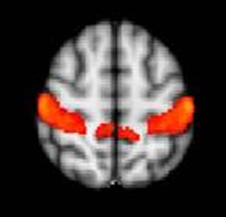
The team found that study participants with ASD and SOR had stronger brain responses to the sensory stimuli in brain areas that process sensory information, the primary somatosensory and auditory cortices, as well as in the amygdala, one of the brain’s emotional centers. The brain responses were even more severe when participants with ASD and SOR were exposed simultaneously to the auditory and tactile stimuli. They also found that all study participants showed a similar initial brain response to the stimuli, but youth with ASD and SOR were much slower in getting used to the stimuli — and reducing their brain response — than were children with ASD but not SOR.
Green said that youth with ASD but not SOR may be compensating through strong brain connectivity between their prefrontal cortex and amygdala, an area involved in attention, response to threat and emotional reactions. The prefrontal cortex, Green said, is helping to regulate the strong response of the amygdala.
‘We don’t have many good treatments for SOR, but our results suggest that an effective treatment might focus on creating coping skills to deal with stimulating environments rather than focusing on changing sensory processing,’ Green said. ‘I also think parents and others working with kids with autism need to be aware of their sensory environments. For example, a child might have more trouble being touched in a loud, crowded room. Or if a child has tactile sensitivity, parents may want to be particularly careful that they are wearing comfortable clothing before trying out a new environment, such as a movie theater or restaurant.’
Going forward, the UCLA team has received a grant from the Simons Foundation to continue to study how youths with ASD and SOR process sensory stimulation. They hope to better characterize the neurobiology of the brain’s reaction.
Funding: This work was supported by grants from the National Institute of Child Health and Human Development, the National Institute of Mental Health, and the National Center for Research Resources, as well as a National Research Service Award predoctoral fellowship (F31 MH093999-01A1).
Source: Kim Irwin – UCLA Health Sciences
Image Credit: The image is credited to UCLA
Original Research: Full open access research for “Neurobiology of Sensory Overresponsivity in Youth With Autism Spectrum Disorders” by Shulamite A. Green, PhD; Leanna Hernandez, MA; Nim Tottenham, PhD; Kate Krasileva, BA; Susan Y. Bookheimer, PhD; and Mirella Dapretto, PhD in JAMA Psychiatry. Published online June 10 2015 doi:10.1001/jamapsychiatry.2015.0737
Abstract
Neurobiology of Sensory Overresponsivity in Youth With Autism Spectrum Disorders
Importance: More than half of youth with autism spectrum disorders (ASDs) have sensory overresponsivity (SOR), an extreme negative reaction to sensory stimuli. However, little is known about the neurobiological basis of SOR, and there are few effective treatments. Understanding whether SOR is due to an initial heightened sensory response or to deficits in regulating emotional reactions to stimuli has important implications for intervention.
Objective: To determine differences in brain responses, habituation, and connectivity during exposure to mildly aversive sensory stimuli in youth with ASDs and SOR compared with youth with ASDs without SOR and compared with typically developing control subjects.
Design, Setting, and Participants: Functional magnetic resonance imaging was used to examine brain responses and habituation to mildly aversive auditory and tactile stimuli in 19 high-functioning youths with ASDs and 19 age- and IQ-matched, typically developing youths (age range, 9-17 years). Brain activity was related to parents’ ratings of children’s SOR symptoms. Functional connectivity between the amygdala and orbitofrontal cortex was compared between ASDs subgroups with and without SOR and typically developing controls without SOR. The study dates were March 2012 through February 2014.
Main Outcomes and Measures: Relative increases in blood oxygen level–dependent signal response across the whole brain and within the amygdala during exposure to sensory stimuli compared with fixation, as well as correlation between blood oxygen level–dependent signal change in the amygdala and orbitofrontal cortex.
Results: The mean age in both groups was 14 years and the majority in both groups (16 of 19 each) were male. Compared with neurotypical control participants, participants with ASDs displayed stronger activation in primary sensory cortices and the amygdala (P < .05, corrected). This activity was positively correlated with SOR symptoms after controlling for anxiety. The ASDs with SOR subgroup had decreased neural habituation to stimuli in sensory cortices and the amygdala compared with groups without SOR. Youth with ASDs without SOR showed a pattern of amygdala downregulation, with negative connectivity between the amygdala and orbitofrontal cortex (thresholded at z > 1.70, P < .05).
Conclusions and Relevance: Results demonstrate that youth with ASDs and SOR show sensorilimbic hyperresponsivity to mildly aversive tactile and auditory stimuli, particularly to multiple modalities presented simultaneously, and show that this hyperresponsivity is due to failure to habituate. In addition, findings suggest that a subset of youth with ASDs can regulate their responses through prefrontal downregulation of amygdala activity. Implications for intervention include minimizing exposure to multiple sensory modalities and building coping strategies for regulating emotional response to stimuli.
“Neurobiology of Sensory Overresponsivity in Youth With Autism Spectrum Disorders” by Shulamite A. Green, PhD; Leanna Hernandez, MA; Nim Tottenham, PhD; Kate Krasileva, BA; Susan Y. Bookheimer, PhD; and Mirella Dapretto, PhD in JAMA Psychiatry. Published online June 10 2015 doi:10.1001/jamapsychiatry.2015.0737


